Image | Statistics | Caption |
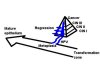 |  | Drawing summarizing the evolution of the transformation zone and the genesis of uterine cervix cancer. |
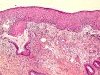
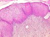
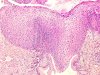
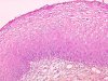
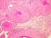
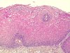
 | | Conization: CIN 1 - condyloma. Lesion situated into the ellipse. Free margins of ectocervix (A,B) and endocervix (C). |
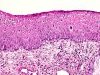
 |  | CIN 1 (flat condyloma): the epithelium organization is disturbed in the lower third. A large abnormal mitotic figure (arrow). |
 | | CIN 1 (flat condyloma): an abnormal mitotic figure (yellow circle), numerous binucleated cells (green circle), superficial parakeratosis (red arrow). Disturbed structural organization of the epithelium in the lower third. |
 | | CIN 1 (flat condyloma): abnormal mitotic figure (green circle), numerous binucleated cells (yellow circle), koilocytotic cells (stars), superficial parakeratosis (red arrow). |
 | | CIN 1 (flat condyloma): two typical koilocytotic cells (green circles) and marked dermal papillae (arrows). |
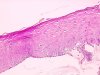
 | | CIN 1 (flat condyloma): occlusion of a glandular orifice. |
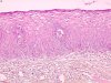
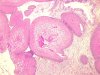
 | | CIN 1 (flat condyloma): flat condyloma (A) and normal ectocervical epithelium (B) next to each other. |
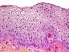
 | | CIN 1 (flat condyloma): koilocytotic cell changes (koilocytosis), superficial parakeratosis and basal cell hyperplasia. |
 | | CIN 1 (flat condyloma): binucleated cells (green circle) and monocellular dyskeratosis (yellow circle). |
 | | CIN 1 with mild koilocytosis, disorganization of the lower third of the epithelium and koilocytes in the upper third. |
 | | CIN 1 with mild koilocytosis, disorganization of the lower third of the epithelium and koilocytes in the upper third. |
 | | CIN 1 with mild koilocytosis, disorganization of the lower third of the epithelium and koilocytes in the upper third. |
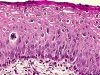
 | | CIN 1: disturbed epithelial maturation with nuclear abnormalities in the lower third of the epithelium. |
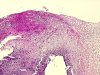
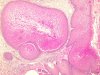
 | | CIN 1 (inverted condyloma): budding aspect of the lesion. |
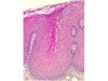
 | | CIN 1 (Inverted condyloma): budding aspect of the lesion (glandular involvement). |
 | | CIN 1 (Inverted condyloma): budding aspect of the lesion (glandular involvement). |
 | | CIN 1 (Inverted condyloma): superficial parakeratosis, mild koilocytosis. |
 | | CIN 1 (Inverted condyloma): mild koilocytosis. |
 | | CIN 1 (Inverted condyloma): disorganization of the lower third of the epithelium, several binucleated cells, koilocytosis. |
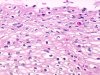
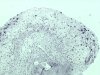
 | | CIN 1 or inflammatory atypia? Disturbed epithelial maturation with nuclear abnormalities in the lower third of the epithelium. Heavy exudate with lymphocytes and polymorphs. Local treatment is needed to allow a specific diagnosis. |
 | | CIN 1 (Flat condyloma): immunohistochemistry, anti-Mib1 antibody: marked positivity of the nuclei of the koilocytotic cells, and of a few nuclei of normal basal cells. Many positive inflammatory cells in the lamina propria. |
 | | CIN 1 (Flat condyloma): in situ hybridization, HPV 16 probe: labelling of the nuclei of numerous superficial cells. |
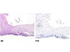 | | HE staining: CIN 1 and then normal ectocervix. Anti-p16 immunolabelling: moderate staining of the CIN 1. No labelling of the normal tissue. |
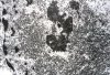 | | CIN 1 (flat condyloma): transmission electron microscopy, koilocytotic cell nucleus with numerous intranuclear viral particles. |
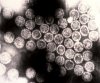 | | CIN 1(flat condyloma), transmission electron microscopy, high magnification of the HPV virus. |
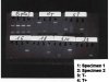 | | HPV typing (PCR): specimen 1: consensus sequences GP5-GP6 and MY11-MY09 present. Specimen 2: negative results. |
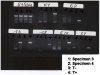 | | HPV typing (PCR): specimen 1: negative results. Specimen 2: HPV 16 positivity (see glossary). |


