Image | Statistics | Caption |
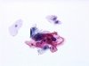
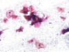
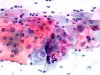
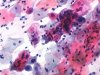
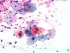
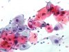
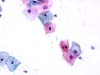
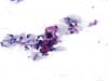
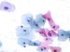
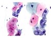
 |  | A and B: LSIL. |
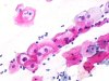 |  | LSIL: eosinophilic squamous cells with a perinuclear empty cavity surrounded by cytoplasmic thickening and with moderate nuclear enlargement: typical koilocytes. (obj. 20x) |
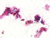
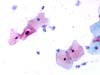
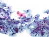
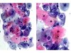
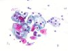 | | LSIL: basophilic and a few eosinophilic squamous cells with a perinuclear empty cavity surrounded by cytoplasmic thickening and with moderate nuclear enlargement: typical koilocytes. (obj. 20x) |
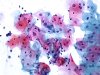
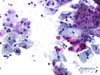
 | | LSIL: basophilic squamous cells with a perinuclear empty cavity surrounded by cytoplasmic thickening and with moderate nuclear enlargement: typical koilocytes. (obj. 20x) |
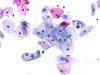
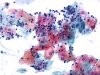
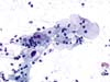
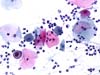
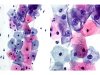
 | | LSIL: eosinophilic squamous cells with dense cytoplasm, parakeratosis and some typical koilocytes. (obj. 20x) |
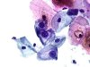
 | | LSIL: group with a typical koilocyte and two parakeratotic cells. (obj. 20x) |
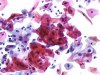
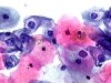
 | | LSIL: typical eosinophilic and basophilic koilocytes associated with some parakeratosis and binucleated cells. (obj. 20x) |
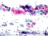
 | | Two koilocytes (arrow) in a field containing some cells with an enlarged nucleus or binucleated with parakeratotic features: LSIL. (obj. 10x) |
 | | LSIL: koilocyte with an enlarged, hyperchromatic nucleus. (obj. 20x) |
 | | LSIL: two eosinophilic koilocytes (arrows) associated with parakeratotic cells. (obj. 20x) |
 | | LSIL: eosinophilic koilocyte (arrow) associated with parakeratotic cells. (obj. 20x) |
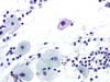
 | | Inflammatory ectocervix: squamous cells with a clear perinuclear halo (arrow) and a nucleus increased in size: Trichomonas vaginalis infection (no visible parasite). No LSIL. (obj. 20x) |
 | | Inflammatory ectocervix: squamous cells with a clear perinuclear halo and a nucleus increased in size: Trichomonas vaginalis infection (no visible parasite). No LSIL. (obj. 20x) |
 | | Squamous cells with an enlarged nucleus, surrounded by an ill-defined clear halo, associated with parakeratotic cells: LSIL. (obj. 20x) |
 | | Squamous cells with an enlarged nucleus, surrounded by an ill-defined clear halo, associated with parakeratotic cells: LSIL. (obj. 20x) |
 | | Squamous cells with an enlarged nucleus, surrounded by an ill-defined clear halo, associated with parakeratotic cells: LSIL. (obj. 20x) |
 | | Squamous cells with an enlarged nucleus, surrounded by an ill-defined clear halo, associated with atypical parakeratotic cells: LSIL. (obj. 20x) |
 | | Typical koilocyte (arrow), eosinophilic cytoplasm, perinuclear empty cavity surrounded by cytoplasmic thickening associated with moderate nuclear enlargement (LSIL). (obj. 10x) |
 | | Typical koilocyte (arrow), eosinophilic cytoplasm, perinuclear empty cavity surrounded by cytoplasmic thickening associated with moderate nuclear enlargement (LSIL). (obj. 20x) |
 | | Typical koilocytes: eosinophilic squamous cells some of them showing a perinuclear empty cavity surrounded by cytoplasmic thickening associated with moderate nuclear enlargement (LSIL). (obj. 20x) |
 | | Typical koilocytes: eosinophilic squamous cells some of them showing a perinuclear empty cavity surrounded by cytoplasmic thickening associated with moderate nuclear enlargement (LSIL). (obj. 20x) |
 | | Typical koilocytes: eosinophilic squamous cells some of them showing a perinuclear empty cavity surrounded by cytoplasmic thickening associated with moderate nuclear enlargement (arrow) (LSIL). (obj. 20x) |
 | | Typical basophilic koilocyte showing a perinuclear empty cavity surrounded by cytoplasmic thickening associated with distinct nuclear enlargement (LSIL). (obj. 20x) |
 | | Typical eosinophilic koilocyte showing a perinuclear empty cavity surrounded by cytoplasmic thickening associated with distinct nuclear enlargement (LSIL). (obj. 20x) |
 | | Typical basophilic koilocyte showing a perinuclear empty cavity surrounded by cytoplasmic thickening and an abnormal nucleus, enlarged and hyperchromatic, with an Indian ink chromatin (arrow) (LSIL). (obj. 20x) |
 | | Typical koilocyte, with eosinophilic cytoplasm showing a perinuclear empty cavity surrounded by cytoplasmic thickening and nuclear enlargement, hyperchromasia, with an Indian ink chromatin (arrow) (LSIL). (obj. 20x) |
 | | Typical basophilic koilocyte showing a double nucleus, accompanied by other slightly atypical cells (LSIL). (obj. 20x) |
 | | Basophilic koilocyte showing an abnormal nucleus (arrow), accompanied with other atypical cells, with eosinophilic cytoplasm and hyperchromatic homogenous nuclei (ellipse) (LSIL). (obj. 20x) |
 | | Atypical cell (arrow): ASC-US (rule out LSIL). (obj. 20x) |
 | | Eosinophilic koilocyte (arrow) showing a perinuclear empty cavity surrounded by cytoplasmic thickening and a bizarre nucleus, enlarged, irregular or double (LSIL). (obj. 20x) |
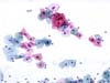
 | | Basophilic koilocyte difficult to detect (arrows) among many intermediate and superficial normal cells (LSIL). (obj. 40x) |
 | | Koilocytotic cell of intermediate type, eosinophilic, showing a perinuclear cytoplasmic clearing and an abnormal nucleus, markedly enlarged and hyperchromatic (LSIL). (obj. 20x) |
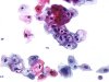
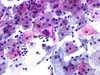
 | | Basophilic koilocytes easily detected among many normal intermediate and superficial squamous cells, in a mildly inflammatory smear (LSIL). (obj. 10x) |
 | | Basophilic koilocytes characterized by their enlarged nuclei and more or less typical perinuclear empty cavity surrounded by cytoplasmic thickening (LSIL). (A: obj. 20x, B: obj. 40x) |
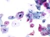
 | | Basophilic koilocytes among many normal intermediate and superficial squamous cells (LSIL). (A: obj. 20x, B: obj. 40x) |
 | | Basophilic koilocytes among many normal intermediate and superficial squamous cells, in a mildly inflammatory smear (LSIL). (A: obj. 20x, B: obj. 40x) |
 | | Basophilic koilocytes and normal intermediate and superficial squamous cells (LSIL). (obj. 40x) |
 | | Pseudokoilocyte: intermediate squamous cell with a normal nucleus and a perinuclear clearing: artefact. (obj. 20x) |
 | | Pseudokoilocyte: the nucleus is pyknotic and there is no optically empty zone. Probably artefact. (obj. 40x) |
 | | Pseudokoilocytes (arrows): intermediate and superficial squamous cells with basophilic or eosinophilic cytoplasm, sometimes of navicular type. There is no evidence of LCIL infection. (obj. 20x) |
 | | Pseudokoilocytes (dotted line): intermediate and superficial cells with basophilic or eosinophilic cytoplasm, normal or with complete cytoplasmic clearing: probable artefact. (obj. 10x) |
 | | Pseudokoilocytes: intermediate and superficial cells with basophilic or eosinophilic cytoplasm, normal or with complete cytoplasmic clearing: probable artefact. (obj. 20x) |
 | | Pseudokoilocytes: intermediate and superficial cells with basophilic or eosinophilic cytoplasm, with complete cytoplasmic clearing without nuclear atypia. Artefact. (obj. 20x) |


