


Learning colposcopy
Colposcopic appearance of normal cervix
The colposcopic examination
Detection of infections & benign conditions of cervix
Detection of cervical neoplasias
Treatment of cervical intraepithelial neoplasia
Treatment by cryotherapy
Treatment by thermal ablation
Treatment by LLETZ (LEEP)
Treatment by cold-knife conization (CKC)
Cases
Normal
Squamous metaplasia and ectropion
Inflammation and cervicitis
Low grade
High grade
Early and advanced cancers
Miscellaneous
Post treatment
Search with IFCPC criteria
Search with Swede score criteria
Quiz Foreword
Acknowledgement
Authors
Suggested citation
Home
Atlas of Colposcopy: Principles and Practice
Filter by language: English / 中文 / Français / Español / Português / Русский
Squamous metaplasia & ectropion / Metaplasia
Go back to the list
Colposcopy report (2011 IFCPC nomenclature):
Swede score:
Final Swede score: 1
Case Summary
Go back to the list
 | |
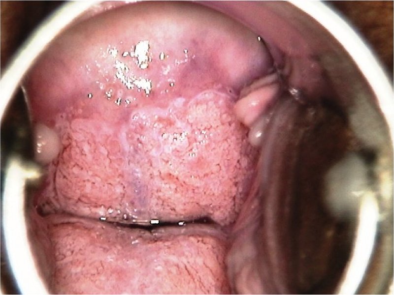
| Speculum examination |
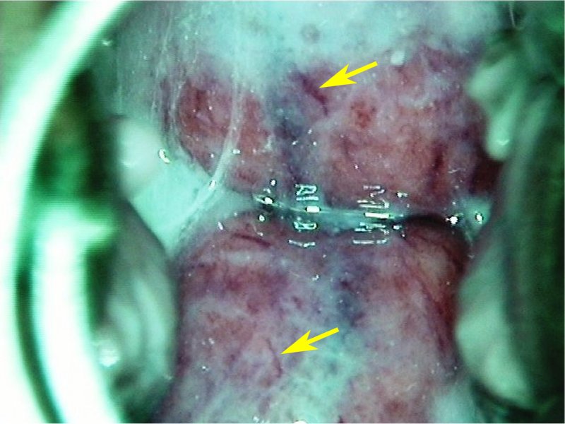 | |
| After normal saline with green filter |
 | |
| After acetic acid |
 | |
| After acetic acid |
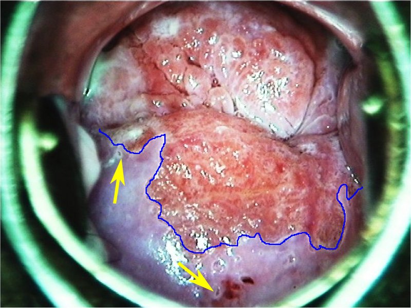 | |
| After acetic acid |
 | |
| After Lugols iodine |
|
|
|
|
|
| |
Swede score:
| Nil or transparent | Thin, milky | Distinct, stearin | |
| Nil or diffuse | Sharp but irregular, jagged, satellites | Sharp and even, difference in level | |
| Fine, regular | Absent | Coarse or atypical vessels | |
| < 5 mm | 5-15 mm or 2 quadrants | >15 mm, 3-4 quadrants, or endocervically undefined | |
| Brown | Faintly or patchy yellow | Distinctly yellow |
Final Swede score: 1
Case Summary
| Provisional diagnosis: | Type 1 transformation zone; normal with ectropion. |
| Management: | Routine screening after 5 years. |
| Histopathology: | Not done. |
| Comment: | Early metaplastic changes are evidenced by the flattening of villi of columnar epithelium and tongue-like inward projection of squamous epithelium. |