


Learning colposcopy
Colposcopic appearance of normal cervix
The colposcopic examination
Detection of infections & benign conditions of cervix
Detection of cervical neoplasias
Treatment of cervical intraepithelial neoplasia
Treatment by cryotherapy
Treatment by thermal ablation
Treatment by LLETZ (LEEP)
Treatment by cold-knife conization (CKC)
Cases
Normal
Squamous metaplasia and ectropion
Inflammation and cervicitis
Low grade
High grade
Early and advanced cancers
Miscellaneous
Post treatment
Search with IFCPC criteria
Search with Swede score criteria
Quiz Foreword
Acknowledgement
Authors
Suggested citation
Home
Atlas of Colposcopy: Principles and Practice
Filter by language: English / 中文 / Français / Español / Português / Русский| Squamous metaplasia Colposcopic features |
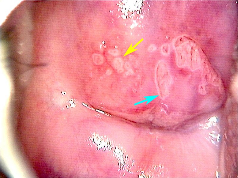
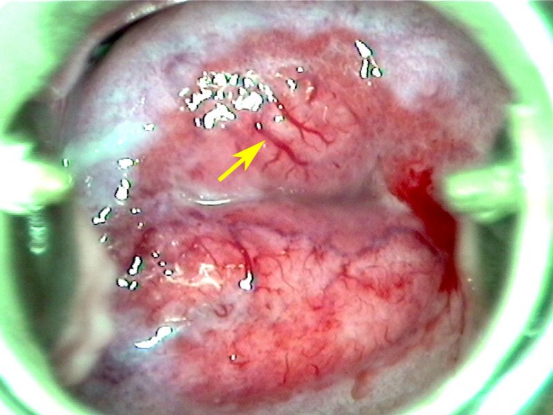
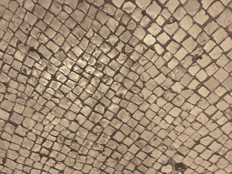
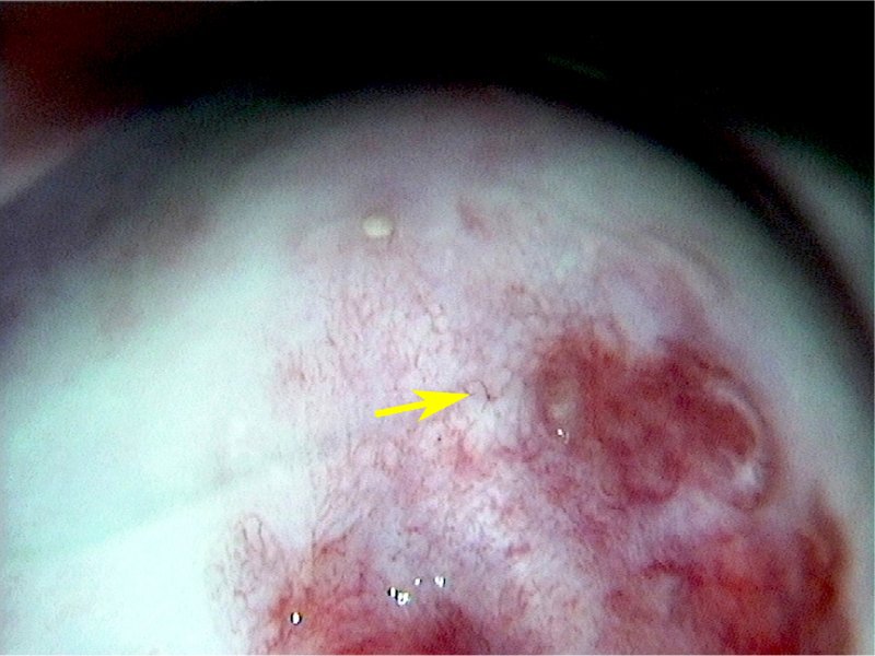
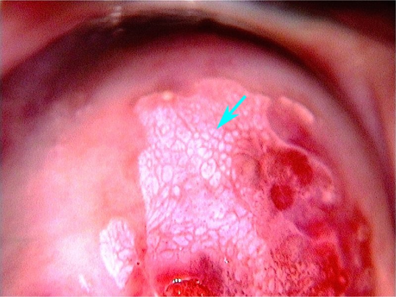
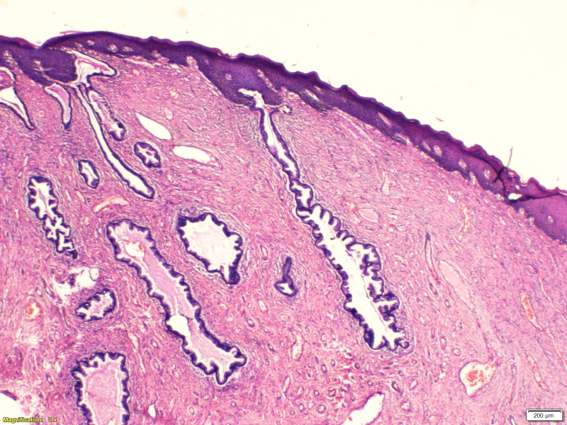
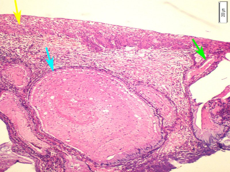
Certain features help to identify the epithelium undergoing squamous metaplasia on colposcopy. Tongue-like projections of the epithelium towards the external os (centripetal extensions) signify that the metaplastic process is still active. These tongue-like projections may be faintly white after application of acetic acid. As the metaplastic epithelium progresses towards the external os, it may cover the glandular crypts originally present in the columnar epithelium or spare them. If the crypts are spared, the patent openings of the crypts are visible as black dots. Sometimes these openings have a thin white margin after application of acetic acid. Crypt openings are also called gland openings. Nabothian cysts (or follicles) are retention cysts formed because of blockage of the crypt openings by the metaplastic epithelium. Nabothian cysts look like white pimples or blisters on the cervix, with fine capillaries of blood vessels stretched over them. As the metaplasia progresses, small patches of columnar epithelium may still remain uncovered. The skipped areas appear as islands of columnar epithelium on the metaplastic epithelium. The normal blood vessels of cervical stroma may be visible through the thin metaplastic epithelium. The vessels have a normal branching pattern, unlike the irregular, abnormal vessels seen on invasive lesions. The stromal capillaries have branches that project towards the surface of the epithelium vertically. The tips of these capillaries may be visible through thin metaplastic epithelium as so-called fine punctations or small red dots. Sometimes the blood vessels are visible as a network of fine capillaries of uniform calibre on the surface. They look like the mosaic tiles on a floor. The mosaic pattern of vessels seen in metaplastic epithelium is known as fine mosaic because of the uniform distribution of the fine capillaries. They are better visualized on an acetowhite background. Metaplastic epithelium may turn white after application of acetic acid (acetowhite). The white area is usually thin, patchy, or transparent. The margin of the white patch is often indistinct, irregular, or fluffy. Uptake of Lugols iodine in the metaplastic epithelium depends on the degree of maturation. In metaplastic epithelium there is usually partial or patchy iodine uptake. With advancing age, the entire metaplastic epithelium (also called immature metaplasia) transforms into mature squamous epithelium, when it may not be possible to distinguish between the original squamous epithelium and the metaplastic squamous epithelium. None of the features of metaplasia will be visible on mature squamous epithelium. Microscopic appearance: Metaplastic epithelium is visible on microscopy as the area between the normal columnar epithelium proximally and the original squamous epithelium distally. The immature epithelium has multiple layers of cuboidal cells with relatively large nuclei, simulating the basal and parabasal cells of the squamous epithelium. Crypt openings and nabothian cysts may be visible. Metaplastic epithelium after complete maturation is indistinguishable microscopically from the original squamous epithelium. |


l.jpg)
a.jpg)

25 avenue Tony Garnier CS 90627 69366, LYON CEDEX 07 France - Tel: +33 (0)4 72 73 84 85
© IARC 2025 - Terms of use - Privacy Policy.
© IARC 2025 - Terms of use - Privacy Policy.