


Learning colposcopy
Colposcopic appearance of normal cervix
The colposcopic examination
Detection of infections & benign conditions of cervix
Detection of cervical neoplasias
Treatment of cervical intraepithelial neoplasia
Treatment by cryotherapy
Treatment by thermal ablation
Treatment by LLETZ (LEEP)
Treatment by cold-knife conization (CKC)
Cases
Normal
Squamous metaplasia and ectropion
Inflammation and cervicitis
Low grade
High grade
Early and advanced cancers
Miscellaneous
Post treatment
Search with IFCPC criteria
Search with Swede score criteria
Quiz Foreword
Acknowledgement
Authors
Suggested citation
Home
Atlas of Colposcopy: Principles and Practice
Filter by language: English / 中文 / Français / Español / Português / Русский| Normal cervical epithelium Squamocolumnar junction |
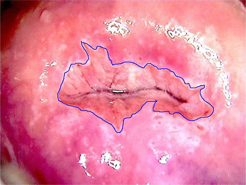
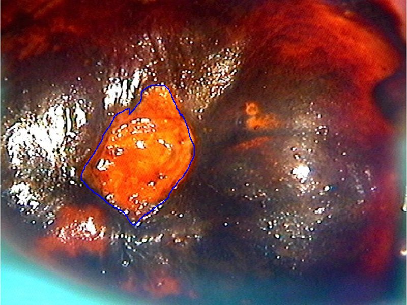
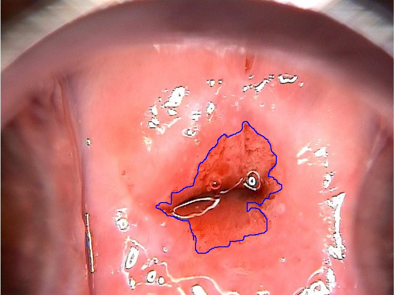
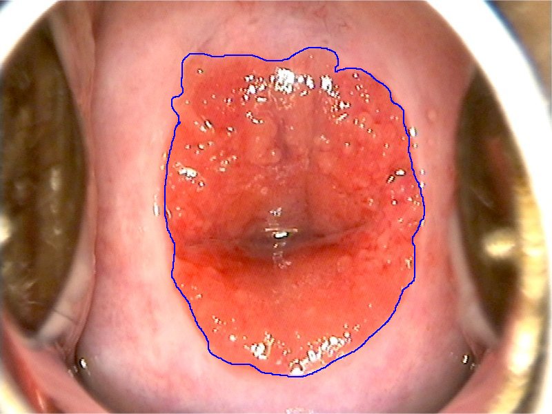
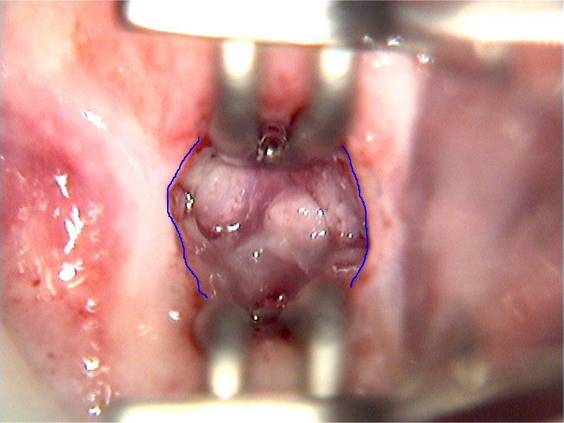
The squamous epithelium and the columnar epithelium meet at a line called the squamocolumnar junction (SCJ). The SCJ is visible as the junction between the pink squamous epithelium and the red columnar epithelium after the cervix is cleaned with normal saline. The SCJ is much better visualized after application of acetic acid as a distinct white line. The junction between the brown squamous epithelium and the red columnar epithelium is also quite evident after application of Lugols iodine. The colposcopist must try to trace the SCJ along its entire course. The SCJ is easily identified if it is at the external os or on the ectocervix. The cervix must be manipulated with an endocervical speculum to visualize the SCJ that extends inside the endocervix.
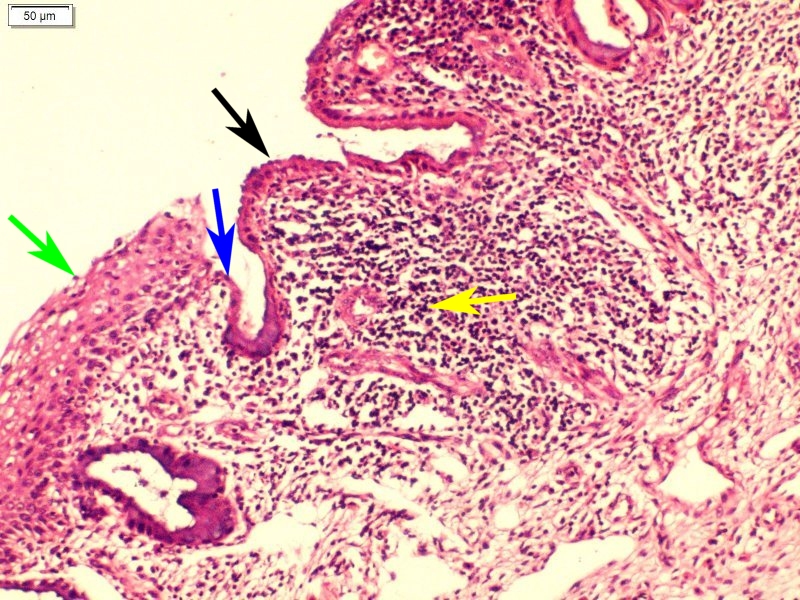
Microscopic features: The junction between the multilayered squamous epithelium and the monolayered columnar epithelium is visible as an abrupt step. The SCJ is prominently seen on colposcopy because of the difference in height between the two epithelia. |


25 avenue Tony Garnier CS 90627 69366, LYON CEDEX 07 France - Tel: +33 (0)4 72 73 84 85
© IARC 2025 - Terms of use - Privacy Policy.
© IARC 2025 - Terms of use - Privacy Policy.