


Learning colposcopy
Colposcopic appearance of normal cervix
The colposcopic examination
Detection of infections & benign conditions of cervix
Detection of cervical neoplasias
Treatment of cervical intraepithelial neoplasia
Treatment by cryotherapy
Treatment by thermal ablation
Treatment by LLETZ (LEEP)
Treatment by cold-knife conization (CKC)
Cases
Normal
Squamous metaplasia and ectropion
Inflammation and cervicitis
Low grade
High grade
Early and advanced cancers
Miscellaneous
Post treatment
Search with IFCPC criteria
Search with Swede score criteria
Quiz Foreword
Acknowledgement
Authors
Suggested citation
Home
Atlas of Colposcopy: Principles and Practice
Filter by language: English / 中文 / Français / Español / Português / Русский| Abnormal colposcopic findings Grade 1 (minor) changes |
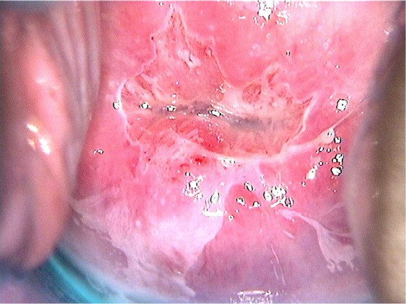
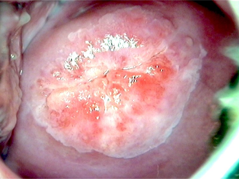
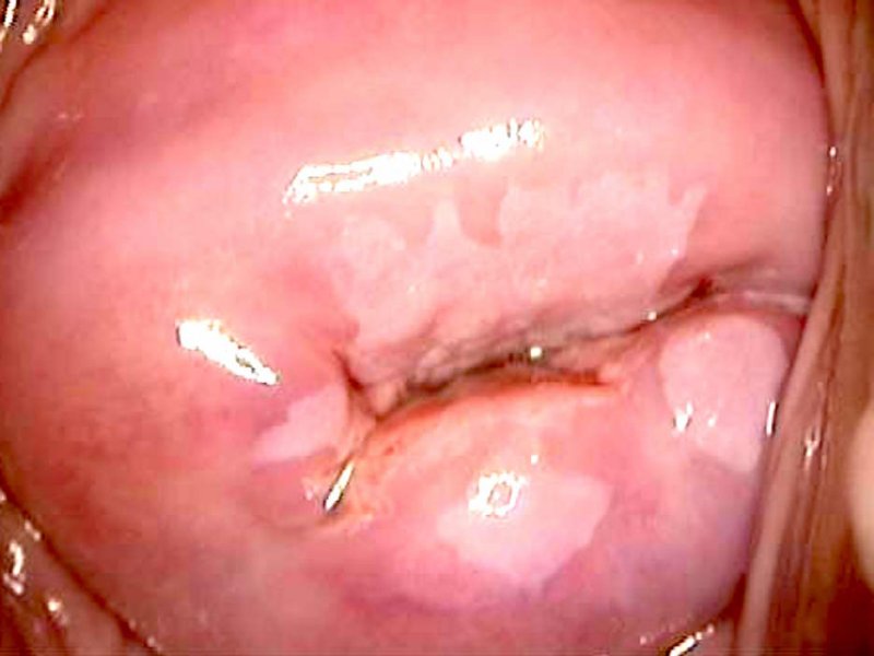
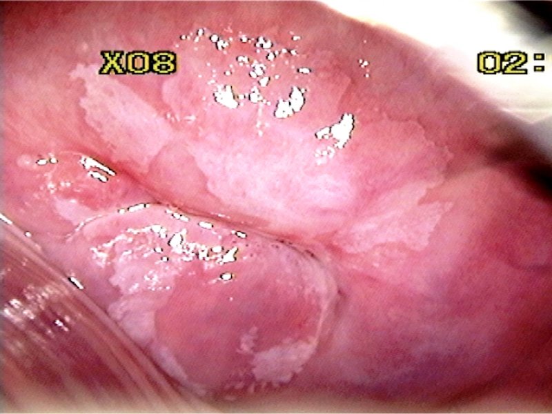
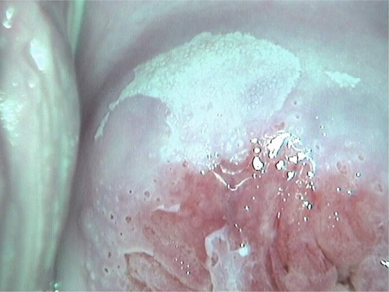
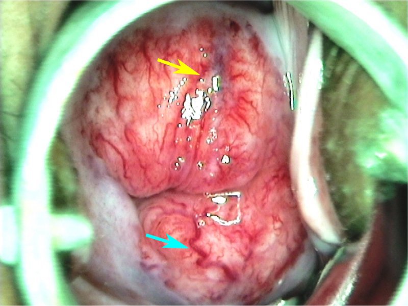
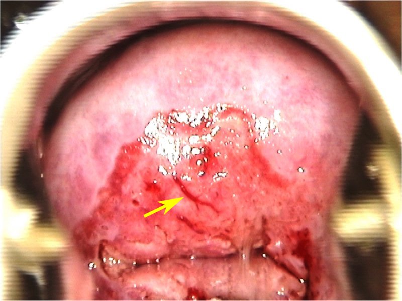
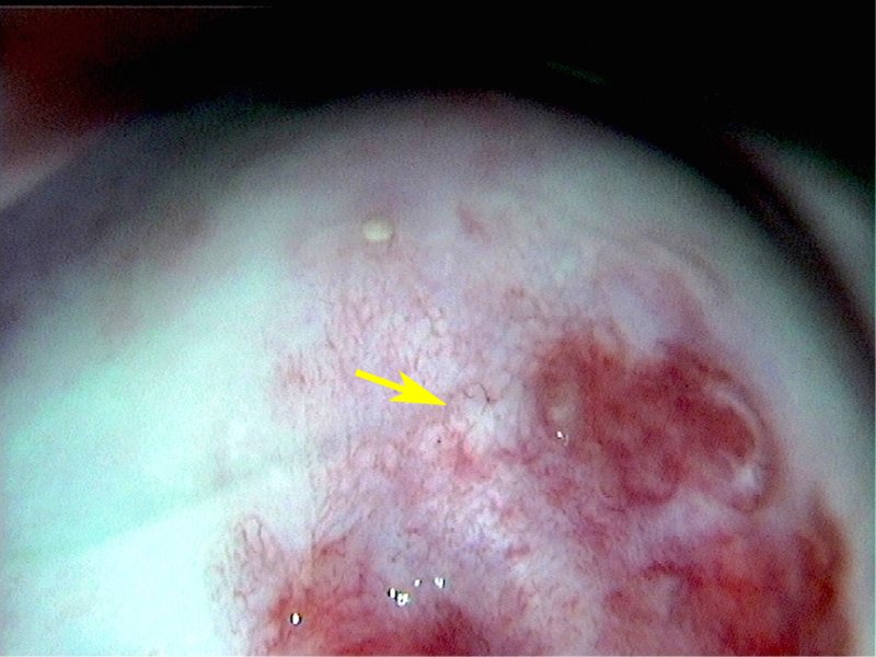
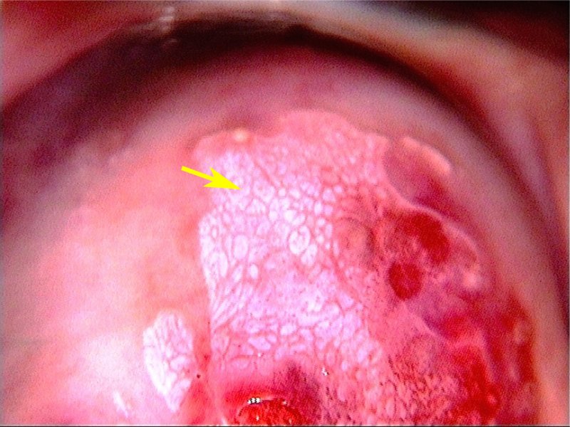
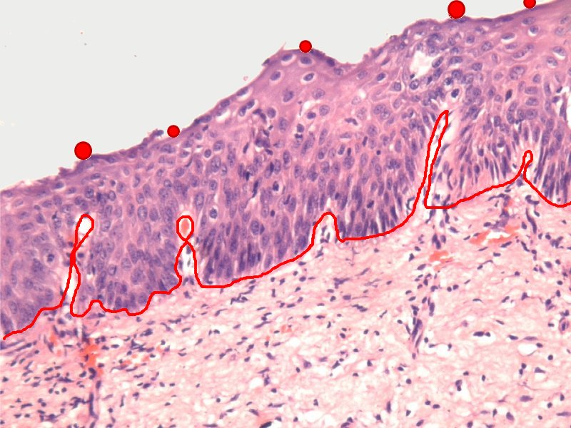
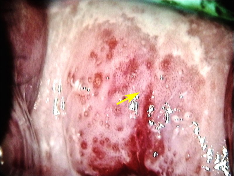
The colposcopically visible abnormalities on the cervix are categorized into grade 1 (minor) changes, grade 2 (major) changes, or non-specific changes. These gradations take into consideration the acetowhite changes, blood vessel patterns (if any), and the changes after application of Lugols iodine. However, acetowhiteness is the most important change to be observed. Grade 1 changes suggest benign conditions or low-grade lesions, whereas grade 2 changes are indicative of high-grade lesions. The non-specific findings are the conditions that may be associated with neoplastic lesions but are also seen in benign conditions. The grade 1 or minor changes may include any of the following features.
Note: The blood vessels constrict because of the influence of acetic acid, and the opaque acetowhite epithelium often hides them. As a result, the blood vessels are no longer seen after application of acetic acid. The blood vessels become visible again when the effect of the acetic acid wears off, after a few minutes. The blood vessels should be assessed (with a green filter) once before application of acetic acid and again a few minutes after application of acetic acid, if there is an acetowhite area. |


25 avenue Tony Garnier CS 90627 69366, LYON CEDEX 07 France - Tel: +33 (0)4 72 73 84 85
© IARC 2025 - Terms of use - Privacy Policy.
© IARC 2025 - Terms of use - Privacy Policy.