Image | Statistics | Caption |
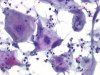 |  | Pap smear from a patient treated with radiotherapy for anal cancer : macrocytosis and macronucleosis, cytoplasmic vacuoles. (obj. 20x) |
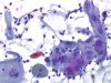 | | Pap smear from a patient treated with radiotherapy for anal cancer : macrocytosis and macronucleosis, prominent nucleoli. (obj. 20x) |
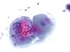 | | Pap smear from a patient treated with radiotherapy for anal cancer : macrocytosis and macronucleosis, cytoplasmic vacuoles and phagocytosis of polymorphs. (obj. 20x) |
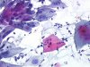 |  | Pap smear from a patient treated with radiotherapy for anal cancer : macrocytosis and macronucleosis, intracytoplasmic inclusion containing a dyskeratotic cell (arrow). (obj. 20x) |
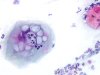 | | Pap smear from a patient treated with radiotherapy for anal cancer : macrocytosis and macronucleosis, cytoplasmic vacuoles and phagocytosis of polymorphs. (obj. 20x) |
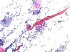 | | Pap smear from a patient treated with radiotherapy for anal cancer: one small and one giant keratinised or necrotic fibre cell. The definite interpretation needs knowing the history. (obj. 20x) |
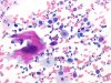 | | Large and bizarre cell (arrow) either in an invasive squamous cell carcinoma (like in this smear), surrounded by typical malignant cells, or after radiotherapy treatment. (obj. 20x) |


