


Introduction
Different approaches to screening and treatment of cervical precancer
Anatomical considerations
Physiological changes of the cervical epithelium
Neoplastic changes of the cervical epithelium
HPV tests Variation between tests
Instruments, consumables, and setup required
Procedure to collect samples for HPV testing
Interpretation of HPV test results
Management of women with a positive HPV test result
Treatment of cervical intraepithelial neoplasia principles
Steps to determine eligibility for ablative treatment
Role of Lugols iodine in identifying the transformation zone for treatment
Treatment by cryotherapy
Treatment by thermal ablation
Using an HPV test as the test of cure in women treated for cervical abnormalities or cervical intraepithelial neoplasia (CIN)
Infection prevention
Case studies
VIA triage outcome (applicable in screen-and-treat setting only) negative cases
VIA triage outcome positive cases
VIA triage outcome suspicious of cancer cases
Foreword
Acknowledgement
Authors
Suggested citation
Copyright
Home / Training / Manuals / Using HPV tests for cervical cancer screening and managing HPV-positive women a practical online guide / Learning
Using HPV tests for cervical cancer screening and managing HPV-positive women a practical online guide
Filter by language: English / Français / EspañolAnatomical considerations Gross anatomy of the cervix | Click on the pictures to magnify and display the legends |
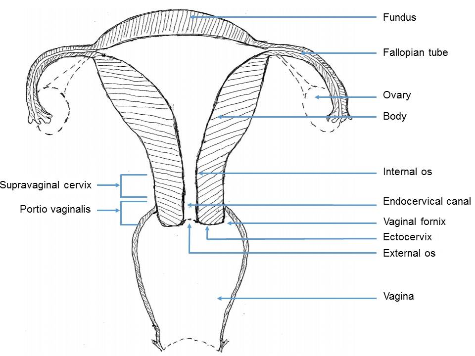
The cervix is the lower part of the uterus and is cylindrical. The length of the cervix is about 34 cm, and its diameter is about 2.5 cm. It has two parts:
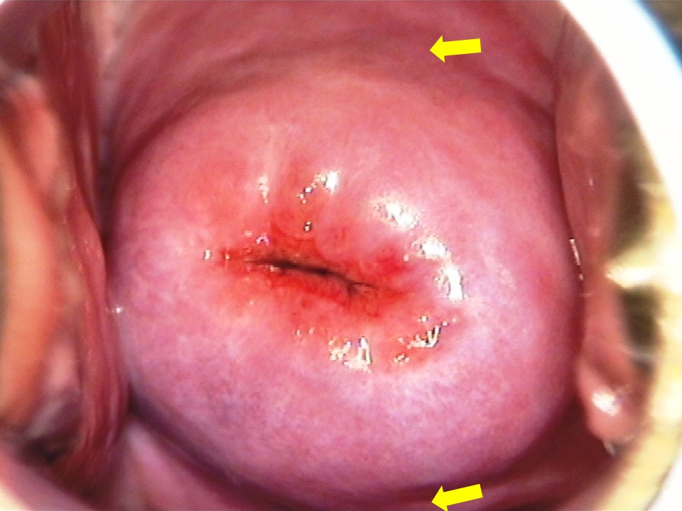
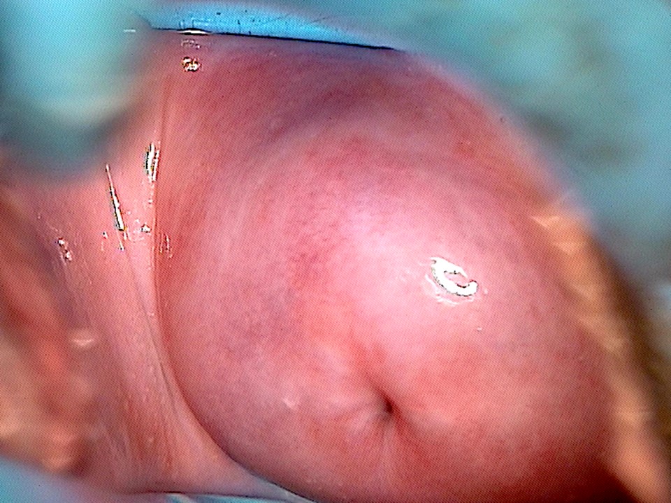
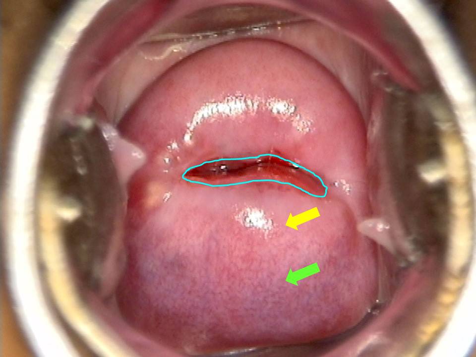
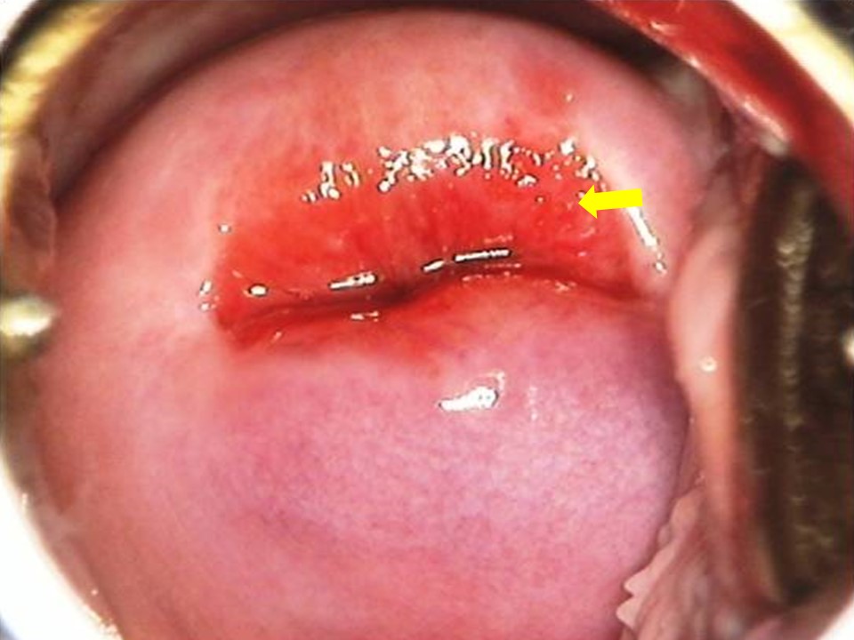
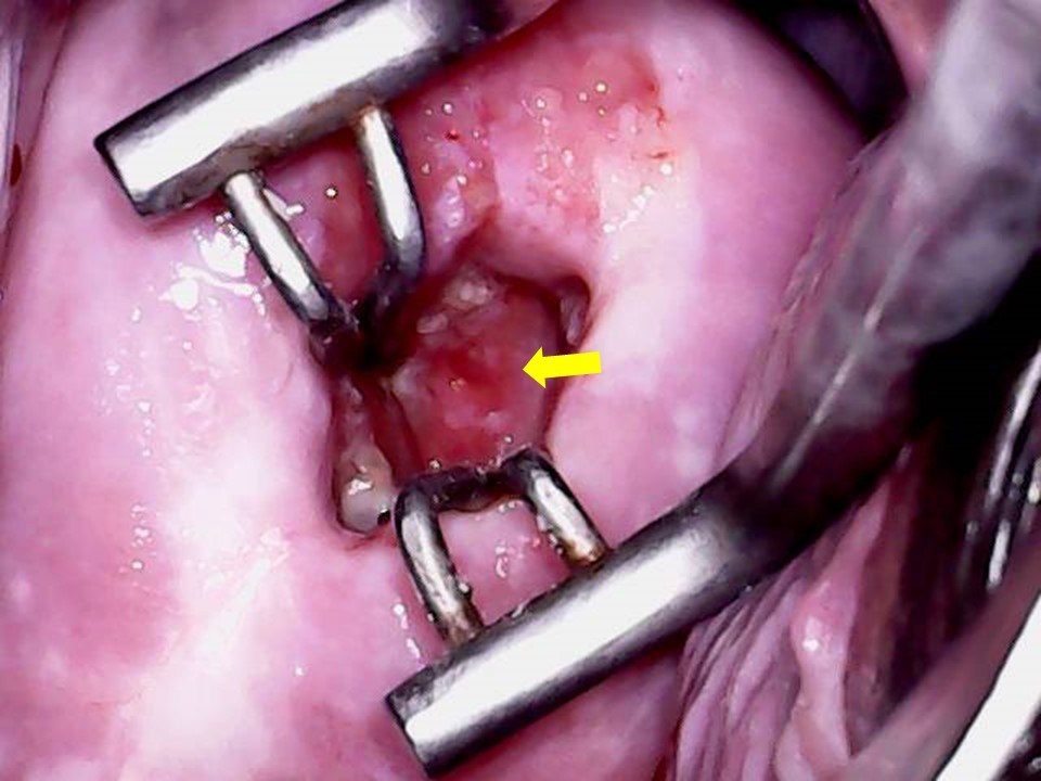
The part of the cervix that is visible during a speculum examination is the ectocervix. The cylindrical canal within the cervix is the endocervix. The endocervix and the ectocervix meet at the external os, which is an opening visible at the centre of the ectocervix. The endocervix joins the uterine cavity through the internal os. The ectocervix has an anterior lip and a posterior lip. In nulliparous women, the external os is small and round and resembles a pinhole. In parous women with history of vaginal childbirth the opening of the ectocervix is wide, it is transversely slit, and its shape resembles that of a fishs mouth. The endocervical canal is a cylindrical space inside the cervix that opens into the vagina through the external os. The upper end of the endocervical canal opens into the body of the uterus through the internal os. The endocervix is lined with columnar epithelium and may not be visualized during a speculum examination of the cervix or during VIA. Endocervical visualization is possible after insertion of endocervical forceps (endocervical speculum). The next section discusses the microscopic anatomy of the cervix. |
IARC, 150 Cours Albert Thomas, 69372 Lyon CEDEX 08, France - Tel: +33 (0)4 72 73 84 85 - Fax: +33 (0)4 72 73 85 75
© IARC 2025 - All Rights Reserved.
© IARC 2025 - All Rights Reserved.