Home / Training / Manuals / A digital manual for the early diagnosis of oral neoplasia / Metastatic tumours

A digital manual for the early diagnosis of oral neoplasia

Metastatic tumours  Search in Medline for Metastatic tumours


Metastatic tumours of the oral cavity are extremely rare. When they do occur, the primary sites implicated are usually breast, lung, kidney, thyroid, colon and prostate. Metastases to the jaw bones are more common from breast and prostate cancer, whereas lung and kidney cancers account for most of the soft tissue metastases Hirshberg A, Shnaiderman-Shapiro A, Kaplan I, Berger R (2007). Metastatic tumours to the oral cavity - Pathogenesis and analysis of 673 cases. Oral Oncol . The mandible, especially the molar area, is more most common affected and attached gingiva is the commonest site of soft tissue metastasis Hirshberg A, Buchner A (1995). Metastatic tumours to the oral region. An overview. Eur J Cancer B Oral Oncol 31B: 355-360.. Pain and swelling may be the presenting symptoms; however, some lesions are asymptomatic and may be an incidental radiographic finding. The majority of these jaw lesions are radiolucent. Sclerotic or mixed density lesions are occasionally seen, usually from breast or prostate cancer Odell EW (2005).  Secondary tumours. In: Barnes L, Eveson,JW, Reichart,P, Sidransky,D eds.  Pathology & Genetics. Head and Neck Tumours, Lyon, IARC Press, 208.. Early manifestation of the gingival metastasis resembles a hyperplastic or reactive lesion, such as a pyogenic granuloma, peripheral giant cell granuloma, or fibrous epulis. The clinical presentation of a metastatic lesion in the oral cavity can be deceiving, leading to a misdiagnosis of a benign process. Therefore, in any case where the clinical presentation is unusual, especially in patients with a known malignant disease imaging, CT and a biopsy is mandatory Hirshberg A, Shnaiderman-Shapiro A, Kaplan I, Berger R (2007). Metastatic tumours to the oral cavity - Pathogenesis and analysis of 673 cases. Oral Oncol . Metastases to the jaws or soft tissues of the oral cavity usually indicate widely disseminated disease and carry poor prognosis; two thirds of patients die in less than one year Odell EW (2005).  Secondary tumours. In: Barnes L, Eveson,JW, Reichart,P, Sidransky,D eds.  Pathology & Genetics. Head and Neck Tumours, Lyon, IARC Press, 208.Hirshberg A, Leibovich P, Horowitz I, Buchner A (1993). Metastatic tumors to postextraction sites. J Oral Maxillofac Surg 51: 1334-1337..

Image

Caption

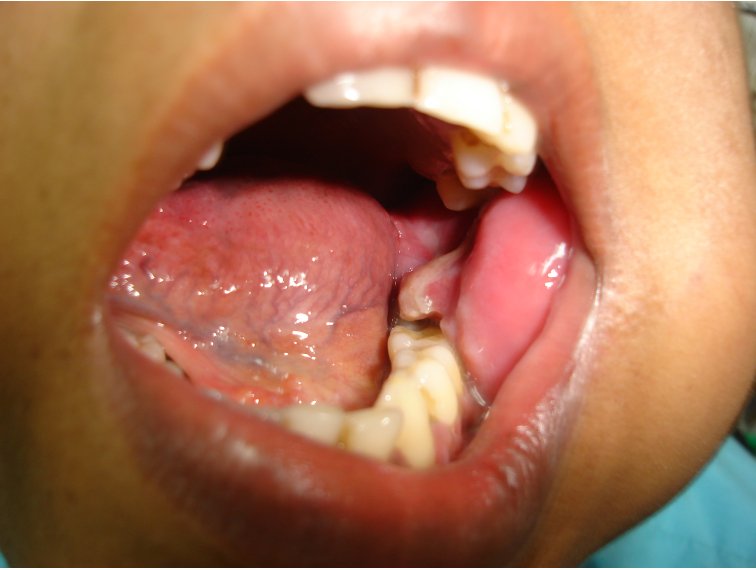
Figure 1: Metastatic tumour in the left lower alveolus. Note the diffuse swelling in the left molar region with a fleshy 2x1 cm pedunculated mass near the last molar. The first two molars are missing.
Figure 2: Panthomograph of the same patient showing a destructive lesion on the left side of the mandible.
Figure 3: CT of the chest of the same patient showing lung carcinoma with enlarged mediastinal and hilar nodes.
Figure 4: Metastatic sarcoma in the left lower gingiva. Note the pedunculated fleshy growth arising from the left lower gingiva in this 44–year-old lady with a retroperitoneal sarcoma.
25 avenue Tony Garnier CS 90627 69366, LYON CEDEX 07 France - Tel: +33 (0)4 72 73 84 85
© IARC 2024 - Terms of use  -  Privacy Policy.