Image | Statistics | Caption |
 |  | Total hysterectomy, macroscopy: relatively well delineated large tumour of the uterine cervix with a grey surface and areas of haemorrhage. |
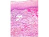 |  | Embryonal rhabdomyosarcoma of the uterine cervix. |
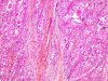 | | Embryonal rhabdomyosarcoma of the uterine cervix with rhabdomyoblasts. |
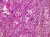 | | Embryonal rhabdomyosarcoma of the uterine cervix with rhabdomyoblasts showing different degrees of differentiation and vacuolisation. |
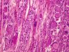 | | Embryonal rhabdomyosarcoma of the uterine cervix with rhabdomyoblasts, different degrees of differentiation and vacuolisation. |
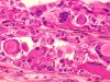 | | Embryonal rhabdomyosarcoma of the uterine cervix with rhabdomyoblasts, different degrees of differentiation and vacuolisation (higher magnification). |
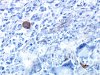
 | | Embryonal rhabdomyosarcoma of the uterine cervix: immuno-histochemistry, anti-vimentin antibody: positive staining of the vascular endothelium and few rhabdomyoblasts. |
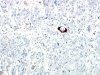
 | | Embryonal rhabdomyosarcoma of the uterine cervix: immuno-histochemistry, anti-smooth muscle actin antibody: positivity of few rhabdomyoblasts. |
 | | Embryonal rhabdomyosarcoma of the uterine cervix: immuno-histochemistry, anti-desmin antibody: positivity of few malignant cells. |


