Image | Statistics | Caption |
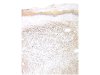
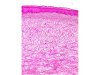 |  | Uterine cervix rhabdomyoma: below an intact squamous epithelium, a mixture of undifferentiated spindle-shaped cells and scattered muscle fibres in various stages of differentiation within a more or less myxoid matrix. |
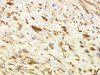
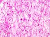 | | Uterine cervix rhabdomyoma: scattered muscle fibres in various stages of differentiation within a more or less myxoid matrix (detail of the previous picture). |
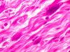 |  | Uterine cervix rhabdomyoma: scattered muscle fibres in various stages of differentiation within a more or less myxoid matrix (detail of the previous picture). |
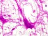 | | Uterine cervix rhabdomyoma: 2 multi-nucleated muscle fibres (arrows) within a myxoid matrix. |
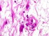 | | Uterine cervix rhabdomyoma: multi-nucleated muscle fibres within a myxoid matrix also containing a vessel. |
 | | Uterine cervix rhabdomyoma: immunohistochemistry, anti-desmin antibody: cytoplasmic positivity of the tumour cells. |
 | | Uterine cervix rhabdomyoma: immunohistochemistry, anti-desmin antibody: strong cytoplasmic positivity of the tumour cells (higher magnification). |
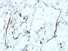 | | Uterine cervix rhabdomyoma: immunohistochemistry, anti-desmin antibody: strong cytoplasmic labelling of the tumour cells (higher magnification). |


