Image | Statistics | Caption |
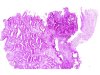
 |  | Conization: microinvasive adenocarcinoma with neoplastic glands seen outside the normal glandular field (for the staging see the TNM and FIGO classifications). |
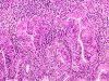
 |  | Microinvasive adenocarcinoma (circle) in the T-zone, 2mm in diameter (for the staging see the TNM and FIGO classifications). |
 | | Microinvasive adenocarcinoma: scattered or clustered neoplastic cells invading the stroma which shows a dense inflammatory infiltrate. |
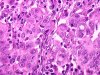 | | Microinvasive adenocarcinoma: scattered or clustered neoplastic cells invading the stroma (higher magnification). |
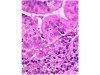 | | Microinvasive adenocarcinoma: invasion of the inflamed connective tissue by scattered neoplastic cells (arrows). |


