Image | Statistics | Caption |
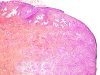
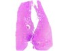 |  | Conization: microinvasive squamous cell carcinoma associated with a carcinoma in situ and a follicular cervicitis (star). Presence of nests of squamous malignant cells (circle) outside the normal glandular area (dots). For staging see the TNM and FIGO classifications. |
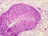
 |  | Microinvasive squamous cell carcinoma. Higher magnification. |
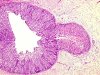 | | Microinvasive squamous cell carcinoma. Higher magnification of the previous picture. Lymphatic emboli. (arrows) |
 | | Micro-invasive squamous cell carcinoma. Higher magnification of the previous picture. Four lymphatic emboli. (arrows) |
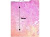 | | Microinvasive carcinoma: mature neoplastic squamous cell nests infiltrating the connective tissue 2mm in depth (bar). |
 | | Microinvasive carcinoma: mature neoplastic squamous cell nests (arrows) infiltrating the connective tissue 2mm in depth, with a carcinoma in situ on the surface (star). |


