Image | Statistics | Caption |
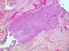
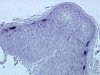
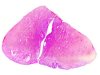 |  | CIN 3: conization, lesion localized between the two arrows. Free resection margins. |
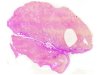 | | CIN 3: conization, lesion localized inside the ellipse, free resection margins. |
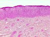 |  | CIN 3: superficial parakeratosis. |
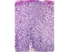
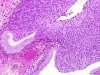
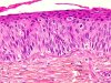 | | CIN 2-3: the cellular organization of the epithelium is disturbed in the lower two-thirds and the cells display a high degree of nuclear and cellular abnormalities with typical and atypical mitoses. |
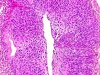
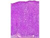 | | CIN 3: the cellular organization of the epithelium is disturbed in more than the lower two-thirds and the cells display a high degree of nuclear and cellular abnormalities with typical and atypical mitoses. |
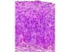 | | CIN 3: the cellular organization is disturbed in almost full thickness of the epithelium and the cells display a high degree of nuclear and cellular abnormalities with typical and atypical mitoses. |
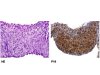
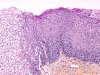 | | CIN 3: superficial parakeratosis. Contrast with the normal exocervical epithelium. |
 | | CIN 3: the cells display a high degree of nuclear and cellular abnormalities in almost full thickness with typical and atypical mitoses and marked superficial parakeratosis. |
 | | CIN 3 (carcinoma in situ): endocervical glandular involvement. |
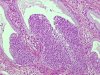 | | CIN 3 (CIS): endocervical glandular involvement. The lesion protrudes into the gland. |
 | | CIN 3 (CIS): endocervical glandular involvement. The lesion protrudes into the gland. |
 | | CIN 3: complete endocervical glandular involvement. |
 | | CIN 3: in situ hybridization - HPV 16 and 18 probes - few positive superficial nuclei. |
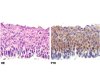 | | HE staining: CIN 3. Immunolabelling with anti-p16: marked positivity of cytoplasm and nuclei of epithelial cells of CIN 3 lesion. |
 | | HE staining: CIN 3. Immunolabeling with anti-p16: heavy labelling of the cytoplasm and nuclei of epithelial of CIN 3 lesion. |


