Image | Statistics | Caption |
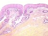
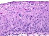
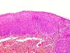
 |  | Conization: CIN 2 (between the two arrows), with involvement of the exocervical margins. |
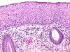
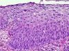
 |  | CIN 1-2: involvement of a glandular orifice. |
 | | CIN 2 with koilocytosis: disorganization of the lower half of the epithelium, cellular criteria of malignancy and viral infection: binucleated cells, abnormal mitotic figures, koilocytes. |
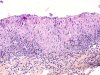 | | CIN 2 with koilocytosis: disorganization of the lower half of the epithelium, cellular criteria of malignancy and viral infection: binucleated cells, abnormal mitotic figures, koilocytes. |
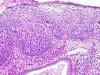 | | CIN 2 with koilocytosis: disorganized architecture of the lower half of the epithelium with cellular criteria of malignancy and viral infection. |
 | | CIN 2 with koilocytosis: disorganized architecture of the lower half of the epithelium with cellular criteria of malignancy and viral infection. |
 | | CIN 2 with koilocytosis: disorganized architecture of the lower half of the epithelium with cellular criteria of malignancy and viral infection. |
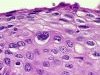 | | CIN 2 with koilocytosis: higher magnification of koilocytes. |
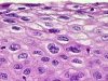 | | CIN 2 with koilocytosis: higher magnification of koilocytes and binucleated cells (arrow). |
 | | CIN 2 with koilocytosis: disturbed maturation of the lower half of the epithelium showing some viral infection cytologically. |
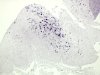 | | CIN 2: in situ hybridization, HPV 31 and 33 probes. A few positive superficial cell nuclei. |
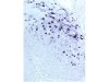 | | CIN 2: in situ hybridization HPV 31 and 33 probes. A few positive superficial cell nuclei. |
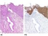 | | HE staining: CIN 2-3. p16 immunolabelling: marked positivity of the cytoplasm and nucleus of dysplastic cells (CIN 2-3). The normal endocervical epithelium is negative. |


