Image | Statistics | Caption |
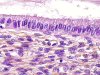 |  | Normal endocervix, epithelium composed of one layer of mucin secreting cells with few ciliated cells (+). |
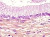 |  | Normal endocervix, epithelium composed of one layer of mucin secreting cells with few reserve cells (arrow). |
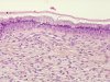 | | Normal endocervix, epithelium composed of one layer of mucin secreting cells with numerous reserve cells. Slight hyperplasia of the reserve cells. |
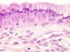 | | Normal endocervix, epithelium composed of one layer of mucin secreting cells with numerous reserve cells. Slight hyperplasia of the reserve cells. |
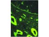 | | Normal endocervix, immunofluorescence - Antibody anti-collagen IV - The epithelium is underlined by a continuous basement membrane (arrow). The basement membranes of the vessels and of the arterial media are also positive (+). |
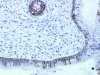
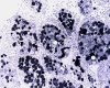
 | | Normal endocervix, immunohistochemistry, anti-Mib1 antibody, labelling of the reserve cell nuclei (circle). |
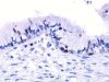
 | | Normal endocervix, immunohistochemistry, antipankeratin (KL1) antibody, partial cytoplasmic labelling of the endocervical cells. |
 | | Distribution of the endocervical glands. (A=External os, B=Isthmus and C= Limits of the glandular field). |
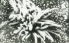 | | Normal endocervix: scanning electron microscopy, one ciliated cell with cilia (7 microns long). |
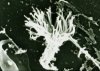 | | Normal endocervix, Cytology, Scanning electron microscopy: one ciliated cell. |
 | | Normal endocervix, transmission electron microscopy, endocervical cells covered with microvilli (green arrows) and with numerous secretory vacuoles (red arrows). |


