Image | Statistics | Caption |
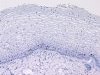
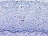
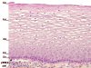 |  | Structure of the ectocervix: CT=connective tissue, BM=basement membrane, L1=basal cells (1 layer), L2=parabasal cells (2 layers), L3=intermediate cells (around 8 layers), L4=superficial cells (5 or 6 layers) and L5=exfoliating cells. |
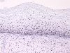
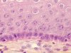 | | Structure of the ectocervix - details of basal, parabasal and intermediate layers: connective tissue, basal cells (one layer), parabasal cells (two layers), intermediate cells (some layers) with inter-cellular bridges. The N/C ratio of basal and parabasal cells is high. |
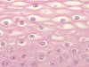 | | Structure of the ectocervix: details of intermediate and superficial layers: intermediate cells (some layers) and superficial cells (5 or 6 layers) with inter-cellular bridges and cytoplasm rich in glycogen. |
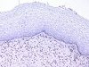
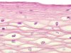 | | Structure of the ectocervix : details of the superficial layers : superficial cells (5 or 6 layers). The N/C ratio is very low and the axis of cells is parallel to the basement membrane. |
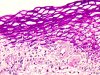 | | Normal ectocervix, PAS staining: intermediate and superficial cells rich in glycogen. |
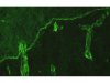 | | Normal ectocervix, immunofluorescence - antibody against collagen IV - The epithelium has a continuous basement membrane (x). The basement membrane of the vessels is also labelled (arrow). |
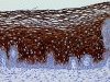 | | Normal ectocervix, immunohistochemistry - antipankeratin (KL1) antibody, positivity of intermediate and superficial cells. Basal and parabasal cells are negative. |
 | | Normal ectocervix, immunohistochemistry: the proliferative activity as visualised by anti-Mib1 antibody is limited to occasional basal and parabasal cells (circles). |
 | | Normal ectocervix: the immunohistochemistry with anti-CD1A antibody shows occasional dendritic cells (circles). |
 | | Normal ectocervix, immunohistochemistry, anti-estrogen receptor antibody, labelling +++ of the nuclei of basal, parabasal, intermediate cells and of fibrocytes of the connective tissue. |
 | | Normal ectocervix, immunohistochemistry, anti-progesteron receptor antibody, nuclei of epithelial cells are not labelled except for some basal cells, but the nuclei of the fibrocytes of the connective tissue are well-stained. |
 | | Normal ectocervix, immunohistochemistry, anti-CD31 antibody, labelling of endothelial cells of the stromal vessels. |
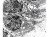
 | | Normal ectocervix - Scanning electron microscopy: superficial cells and Döderlein bacilli. |
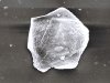 | | Normal ectocervix, superficial cell, scanning electron microscopy: flat cell with a small nucleus (arrow). |
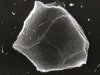 | | Normal ectocervix, superficial cell, scanning electron microscopy: Flat cell with traces of other cells and several Döderlein bacilli. |
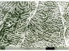 | | Normal ectocervix, superficial cell, scanning electron microscopy (high magnification): intermediate cell surface with numerous ridges (hemi-desmosomes?). |
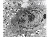
 | | Normal ectocervix, transmission electron microscopy: one basal cell with hemi-desmosomes (HD) on lamina densa (LD) and the connective tissue (CT). Mx14000 |
 | | Normal ectocervix, transmission electron microscopy: one intermediate cell with desmosomes (D), keratinosomes (K) and clusters of glycogen (G). Mx17000 |


