Image | Statistics | Caption |
![](pic/shc000002.jpg) | ![Statistics are not available](public/images/icon/statsbw.png) | 
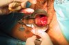
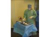
Preparation of sterile material before conization. |
![](pic/shc000004.jpg) | ![Statistics are not available](public/images/icon/statsbw.png) | 
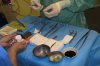
Preparation of the material before conization: antiseptic, acetic acid (5%), Lugol's iodine. Surgical material: forceps, cold knife, scissors, forceps (Pozzi), dilator (n°4 Hegar candle), needle holder, aseptic forceps, electric knife. |
![](pic/shc000005.jpg) | ![Statistics are not available](public/images/icon/statsbw.png) | 
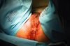
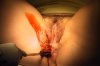
Disinfection of the area with iodine. |
![](pic/shc000006.jpg) | ![Statistics are not available](public/images/icon/statsbw.png) | Preliminary urinary bladder catheterization. |
![](pic/shc000007.jpg) | ![Statistics are not available](public/images/icon/statsbw.png) | Draping of the operating field. |
![](pic/shc000008.jpg) | ![Statistics are not available](public/images/icon/statsbw.png) | 
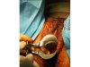
Insertion of the speculum. |
![](pic/shc000009.jpg) | ![Statistics are not available](public/images/icon/statsbw.png) | Anesthesia of the uterine cervix (1% adrenalinated xylocaine). |
![](pic/shc000012.jpg) | ![Statistics are not available](public/images/icon/statsbw.png) | 
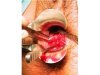
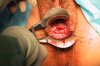
Uterine cervix before conization. |
![](pic/shc000013.jpg) | ![Statistics are not available](public/images/icon/statsbw.png) | 
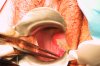
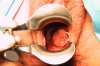
Uterine cervix after application of Lugol's iodine, before the conization. |
![](pic/shc000015.jpg) | ![Statistics are not available](public/images/icon/statsbw.png) | 
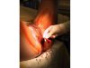
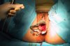
Cold knife conization. Circular incision delineating the lesion. |
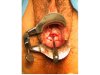
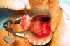
![](pic/shc000018.jpg) | ![Statistics are not available](public/images/icon/statsbw.png) | Cone removal. |
![](pic/shc000021.jpg) | ![Statistics are not available](public/images/icon/statsbw.png) | 
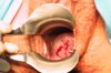
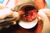
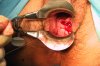
Uterine cervix after cold knife conization. Dilator(Hegar candle) in place in the endocervix. |
![](pic/shc000024.jpg) | ![Statistics are not available](public/images/icon/statsbw.png) | Suture of the uterine cervix and realisation of the first Stumdorf stitch on the anterior lip of the uterine cervix. |
![](pic/shc000026.jpg) | ![Statistics are not available](public/images/icon/statsbw.png) | Suture (Stumdorf stitch) of the anterior lip of the uterine cervix. |
![](pic/shc000031.jpg) | ![Statistics are not available](public/images/icon/statsbw.png) | Suture (Stumdorf stitch) of the posterior lip of the uterine cervix. |
![](pic/shc000032.jpg) | ![Statistics are not available](public/images/icon/statsbw.png) | Tying up the posterior suture (Stumdorf stitch). |
![](pic/shc000033.jpg) | ![Statistics are not available](public/images/icon/statsbw.png) | 
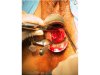
Final appearance of the uterine cervix after tying up the two sutures (Stumdorf stitches) and before carrying out the corner stitch. |
![](pic/shc000039.jpg) | ![Statistics are not available](public/images/icon/statsbw.png) | Cervix suture. |
![](pic/shc000042.jpg) | ![Statistics are not available](public/images/icon/statsbw.png) | Final appearance of the uterine cervix. Permeable cervix with a dilator (n°4 Hegar candle). |