English
Français
Home
Research projects
Breast cancer
Cancer control
Cervical cancer
Colorectal cancer
Epidemiology
HPV vaccine
Oral cancer
Training materials
Manuals
eLearning courses
Digital learning series
Videos tutorials
Other useful Screening videos
Audio presentations
Quick clinical reference charts
Online Library
IARC Publications
Scientific papers
Collaborators
About the group
Visitors and News
Menu
Home
Research projects
Cervical cancer
Breast cancer
Oral cancer
HPV vaccine
Training materials
Manuals
eLearning courses
Digital learning series
Videos
Other useful Screening videos
Audio presentations
Quick clinical reference charts
Online Library
IARC Publications
Scientific papers
Collaborators
About the group
Visitors and News
Histopathology Atlas
Cytopathology Atlas
Oral Atlas
Foreword
Glossary definition
Glossary abbreviation
Atlas contributors
Copyright
Classifications
TNM/FIGO
WHO
Bethesda 2001
References and web links
Home
/
Training
/
Manuals
/
Histopathology of the uterine cervix - digital atlas
/ Micro anatomy of the uterine cervix
Histopathology of the uterine cervix - digital atlas
Micro anatomy of the uterine cervix
Filter by language:
English
/
Français
/
Portugues
/
中文
-
Go back to the Histopathology list
Go back to the Cytopathology list
Foreword
Glossary definition
Glossary abbreviation
Atlas contributors
Copyright
Classifications
TNM/FIGO
WHO
Bethesda 2001
Diagnostic problems and pitfalls
References and web links
Histopathology - Beginner
Histopathology - Advanced
Cytopathology - Beginner
Cytopathology - Advanced
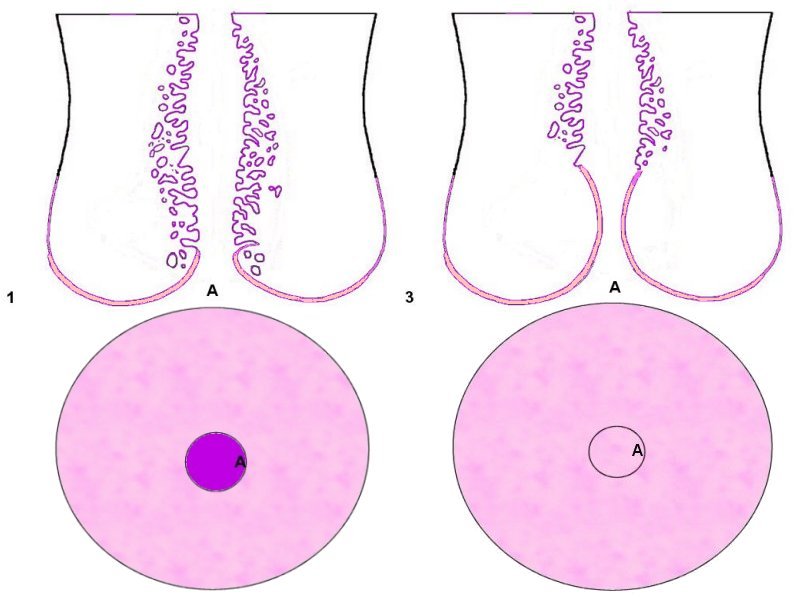
Micro anatomy of the uterine cervix: 1 = Nulliparous, 3 = Menopausal (A: External os, violet area = non-keratinized squamous epithelium, purple area = glandular epithelium composed of one layer of mucin secreting and ciliated cells).
Comments:
Write an online comment about this picture
and share your thoughts with other readers.
25 avenue Tony Garnier CS 90627 69366, LYON CEDEX 07 France - Tel: +33 (0)4 72 73 84 85
© IARC 2024 -
Terms of use
-
Privacy Policy
.