Home / Training / Manuals / Histopathology of the uterine cervix - digital atlas / Glossary Definitions
Histopathology of the uterine cervix - digital atlas
Glossary Definitions
Filter by language: English / Français / Portugues / 中文|
|
 |
|
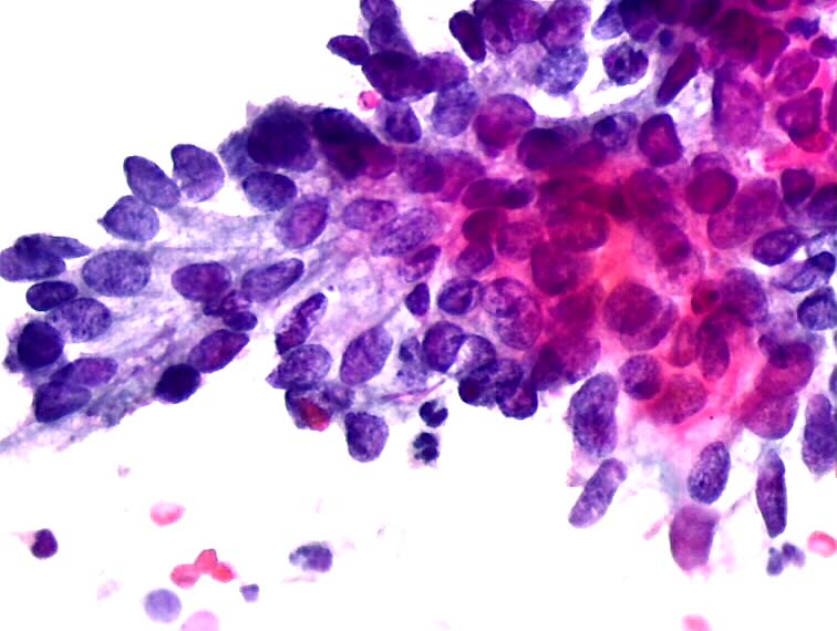
In situ adenocarcinoma Lesion in which, histologically, the normal epithelium of glands is replaced totally or partially by malignant glandular epithelium. The malignant cells do not invade the stroma. In cytology, The Bethesda 2001 System has individualized in situ adenocarcinoma (AIS) from atypia of glandular cells (AGC).Diagnostic criteria of AIS are reproducible and predictive (but remember that HSIL can mimic AIS). Cells are mostly in aggregates, less frequently isolated. Strips of epithelium with altered nuclear polarity, luminal border; with or without stratification and large tissue fragments with branching pattern and gland openings are seen (rosettes or acinar pattern). Nuclei are crowded with lack of cytoplasmic borders and no honeycomb arrangement. Cells at the periphery show feathery pattern. Nuclei are minimally enlarged (8-15 microns), round to oval, hyperchromatic, with granular chromatin. Nucleoli may or may not be present and mitoses are rare. Cytoplasm is scant. Background is clean. Histopathology atlas Cytopathology atlas |
25 avenue Tony Garnier CS 90627 69366, LYON CEDEX 07 France - Tel: +33 (0)4 72 73 84 85
© IARC 2025 - Terms of use - Privacy Policy.
© IARC 2025 - Terms of use - Privacy Policy.


