English
Français
Home
Research projects
Breast cancer
Cancer control
Cervical cancer
Colorectal cancer
Epidemiology
HPV vaccine
Oral cancer
Training materials
Manuals
eLearning courses
Digital learning series
Videos tutorials
Other useful Screening videos
Audio presentations
Quick clinical reference charts
Online Library
IARC Publications
Scientific papers
Collaborators
About the group
Visitors and News
Menu
Home
Research projects
Cervical cancer
Breast cancer
Oral cancer
HPV vaccine
Training materials
Manuals
eLearning courses
Digital learning series
Videos
Other useful Screening videos
Audio presentations
Quick clinical reference charts
Online Library
IARC Publications
Scientific papers
Collaborators
About the group
Visitors and News
Histopathology Atlas
Cytopathology Atlas
Oral Atlas
Foreword
Glossary definition
Glossary abbreviation
Atlas contributors
Copyright
Classifications
TNM/FIGO
WHO
Bethesda 2001
References and web links
Home
/
Training
/
Manuals
/
Histopathology of the uterine cervix - digital atlas
/ Glossary Definitions
Histopathology of the uterine cervix - digital atlas
Glossary Definitions
Filter by language:
English
/
Français
/
Portugues
/
中文
-
Go back to the Histopathology list
Go back to the Cytopathology list
Foreword
Glossary definition
Glossary abbreviation
Atlas contributors
Copyright
Classifications
TNM/FIGO
WHO
Bethesda 2001
Diagnostic problems and pitfalls
References and web links
Histopathology - Beginner
Histopathology - Advanced
Cytopathology - Beginner
Cytopathology - Advanced
Abnormalities of glandular cells
Abnormalities of squamous cells
Acanthosis
Acetic acid test
Actinomyces
Actinomycosis
Adenocarcinoma
Adenofibroma
Adenosis
Adenosquamous carcinoma
AGC: atypical glandular cells
Amphophilic
Anisokaryosis
ASC: atypia of squamous cells
ASC-H
ASC-H and atrophy
ASC-H and atypical immature metaplasia
ASC-H and reserve cell hyperplasia
ASC-US
ASC-US in metaplasia or atrophy
ASC-US on large squamous cells
Atrophic cervix
Atrophic menopausal smear (atrophic pattern)
Atrophy
Atypia of repair
Atypical cells
Bacterial vaginosis
Basal cell hyperplasia
Benign cellular modifications
Bethesda
Bilharziasis of the uterine cervix
Buschke-Lowenstein tumour
Carcinoma in situ
Cell-in-cell image
Cervicitis
Chlamydiae
CIN 1
CIN 2
CIN 3
Clear cell adenocarcinoma
Clue cells
Condyloma
Condyloma acuminatum
Conization
Cytobrush
Dark nuclei
Deciduosis
Depth of invasion
Diethylstilbestrol (DES)
Dyskeratotic
Dystrophy
Early stromal invasion
Embryonal rhabdomyosarcoma
Endocervical polyp
Endocervicitis
Endometrial adenocarcinoma
Endometriosis
Eosinophilic
Exo and endocervicitis
Exocervicitis
Flat condyloma
Florid ectopy
Follicular cervicitis
Gardnerella vaginalis
Glandular atypia
Glandular dysplasia
Glycogen
Granulation tissue
Herpes
High-grade squamous intraepithelial lesion (HSIL)
Histiocytes and small cells
Histiocytic
HPV
HPV changes: binucleation
HPV changes: parakeratosis and dyskeratosis
HPV induced changes: koilocytosis
HPV testing
HPV typing
HSIL with features of moderate dysplasia
HSIL with features of severe dysplasia - CIS
HSIL with features suspicious for invasion
Hybrid capture
Hyperchromatic
Hyperplasia
Hysterectomy
Immature squamous metaplasia
In situ adenocarcinoma
In situ hybridization
Inadequate biopsy
Inflammation
Inflammatory atypias
Invasive adenocarcinoma
Invasive squamous cell carcinoma
Invasive verrucous carcinoma
Inverted condyloma
IUD
IUD associated cytological changes
Keratinized
Koilocyte
Koilocytosis
Langerhan's cells
Langhans cells
LEEP
Leiomyosarcoma
Leukoplakia
Low-grade squamous intraepithelial lesion (LSIL)
Luteal pattern
Macrophage
Malignant lymphoma
Mature squamous metaplasia
Metaplasia
Microinvasive adenocarcinoma
Microinvasive squamous cell carcinoma
Minimal deviation adenocarcinoma
Mullerian papilloma
Mycosis
Nabothian cyst
Necrotic
Non-specific inflammation
Normal ectocervix
Normal endocervical cells
Normal endocervix
Nuclear hypertrophy
Oestrogenic pattern
Osseous and cartilaginous metaplasia
p16ink4a
Papillary adenofibroma
Papillary serous adenocarcinoma
Parakeratosis
Parasites
PCR typing of HPV
Peutz-Jeghers syndrome
Pinworm ovum
Pleomorphic
Pseudo-eosinophilia
Pyogenic granuloma
Radiation-induced changes
Reactive cellular changes
Repair
Reserve cells
Rhabdomyoma
Rosette-like groups
Schiller test
Specific inflammations
Spongiosis
Squames
Squamo-columnar junction
Squamous atypia
Squamous metaplasia
Squamous papilloma
Tingible (bodies)
TNM
Trachelectomy
Transformation zone
Transitional metaplasia
Trichomonas vaginalis
Tubal metaplasia
Tuberculosis
Warty squamous cell carcinoma
Well differentiated invasive adenocarcinoma
Wuchereria bancrofti
labial mucosa
lingual tissue
pan
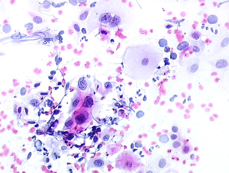
Anisokaryosis
This is one of the main features of cancer cells: variation of nuclear size and shape from cell to cell. Referred to as nuclear pleomorphism and anisonucleosis.
Cytopathology atlas
25 avenue Tony Garnier CS 90627 69366, LYON CEDEX 07 France - Tel: +33 (0)4 72 73 84 85
© IARC 2024 -
Terms of use
-
Privacy Policy
.