Image | Statistics | Caption |
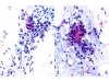
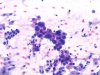
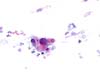
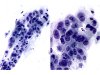
 |  | Inflammatory and bloody smear containing atypical glandular cells preserving their columnar shape with enlarged nuclei (arrows). AGC. (A and B: obj. 20x) |
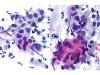
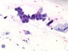
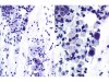
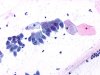
 | | Smear from the transformation zone and endocervix: sheets of atypical glandular cells with enlarged nuclei with similar chromatin pattern in all cells. AGC. (A and B: obj. 20x) |
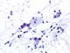
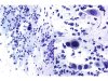
 | | Sheet of atypical glandular cells with enlarged nuclei with similar chromatin pattern in all cells. Compare with some normal columnar cells in the vicinity (dotted line). AGC. (obj. 40x) |
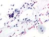
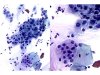
 | | High magnification of atypical columnar cells with enlarged nuclei but with similar chromatin pattern in all cells. No visible nucleolus. AGC. (A and B: obj. 40x) |
 |  | Endocervical smear: sheet of atypical cells of glandular type with enlarged nuclei and a regular chromatin pattern. Compare with normal columnar cells. Nucleoli are occasionally visible. AGC. (obj. 20x) |
 | | Endocervical smear: sheet of atypical cells of glandular type with enlarged nuclei and a regular chromatin pattern. Compare with normal columnar cells. Nucleoli are occasionally visible. AGC. (obj. 20x) |
 | | Endocervical smear: sheet of atypical cells of glandular type with enlarged nuclei and a regular chromatin pattern. Compare with normal columnar cells. Nucleoli are occasionally visible. AGC. (obj. 20x) |
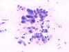
 | | Atypical cells, glandular with regular outlines and homogenous chromatin. AGC. (obj. 20x) |
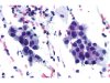
 | | Cluster of atypical cells of glandular origin: enlarged nuclei with homogenous and dense chromatin pattern. AGC. (obj. 20x) |
 | | Three endocervical glandular cells, one of them is atypical with an enlarged nucleus, with regular outlines and homogenous chromatin. AGC. (obj. 20x) |
 | | High magnification of two isolated atypical cells of metaplastic or glandular type with enlarged nuclei showing a similar chromatin pattern. Compare with normal columnar cells. ASC-H. (A: obj. 20x, B: obj. 40x) |
 | | Isolated atypical cells of glandular or metaplastic type with enlarged nuclei showing a similar chromatin pattern. Compare with normal columnar cells. ASC-H. (A: obj. 20x, B: obj. 40x) |
 | | AGC: sheets of slightly atypical endocervical glandular cells with slightly enlarged nuclei (N/C not much disturbed), with irregular outlines but homogenous chromatin pattern. Compare with normal counterparts. (A: obj. 20x, B: obj. 40x) |
 | | AGC: sheet of slightly atypical metaplastic or endocervical glandular cells with slightly enlarged nuclei (N/C not much disturbed), with irregular outlines but homogenous chromatin pattern. Compare with normal counterparts. (A: obj. 20x, B: obj. 40x) |
 | | Well-differentiated endocervical adenocarcinoma. Aspect of the smear: AGC, favor neoplastic. Columnar cells in a palisading arrangements with regular and nucleolated nuclei which are enlarged (compare with squamous cells). (obj. 20x) |


