Image | Statistics | Caption |
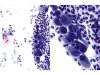
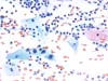 |  | Parabasal cells with nuclear enlargement, irregular nuclear outlines and coarse chromatin (hyperchromatic nucleus (arrow) and binucleation). No koilocyte . LSIL. (obj. 20x) |
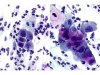
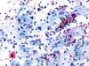 |  | Parabasal cells with marked nuclear enlargement, with hyperchromatic and homogenous chromatin (black arrow): LSIL. The presence of basal atypical cell (purple arrow): HSIL. (obj. 20x) |
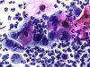
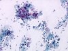 | | Intermediate and parabasal cells with nuclear enlargement, irregular nuclear outlines. HSIL. (obj. 10x) |
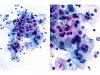 | | Intermediate cells with nuclear enlargement, irregular nuclear outlines and coarse chromatin. HSIL. (A: obj. 20x, B:obj. 40x) |
 | | Parabasal cells with enlarged nuclei, with irregular outlines in an inflammatory smear: HSIL. (A: obj. 20x, B: obj. 40x) |
 | | Inflammatory smear with parabasal cells with enlarged nuclei and irregular outlines: HSIL. (obj. 40x) |
 | | Inflammatory smear with intermediate or parabasal cells with an enlarged nucleus, with irregular outlines: HSIL. (obj. 40x) |


