Image | Statistics | Caption |
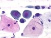
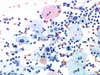
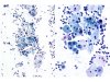
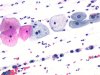 |  | Smear containing basal cells, arranged in Indian files, with enlarged and irregular nuclei and a dense chromatin, suggesting high grade SIL: ASC-H vs HSIL. (obj. 20x) |
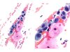
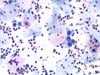
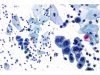
 |  | Smear containing isolated basal cells, with enlarged and irregular nuclei and a dense chromatin, suggesting high grade SIL: ASC-H vs HSIL. (A: obj. 20x, B: obj. 40x) |
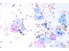
 | | Smear containing isolated basal cells, with enlarged and irregular nuclei (arrow) and a dense chromatin, suggesting high grade SIL: ASC-H vs HSIL. (A: obj. 20x, B: obj. 40x) |
 | | Smear containing basal cells, with enlarged, irregular nuclei and dense chromatin (arrow), suggesting a high grade SIL: ASC-H vs HSIL. (obj. 40x) |
 | | Smear containing one basal cell (arrow) with an enlarged, irregular nucleus and dense chromatin, suggesting a high grade SIL: ASC-H vs HSIL. (obj. 40x) |
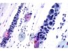
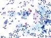
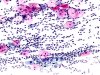 | | Inflammatory smear containing parabasal cells with enlarged nuclei, with irregular outlines surrounded by a clear halo, grouped in line: ASC-H (HSIL?) (obj. 10x) |
 | | Cluster of basal cells and some isolated cells with enlarged nuclei, an irregular chromatin and a thickened nuclear membrane. ASC-H. (A, B, C and D : obj. 40x) |
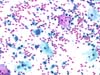
 | | Syncytial group of basal and parabasal cells with enlarged nuclei (immature metaplasia), an irregular but clear chromatin and a thickened nuclear membrane. ASC-H (HSIL?). (A: obj. 20x, B: obj. 40x) |
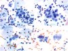
 | | Isolated atypical cells, of intermediate or parabasal type, with nuclear enlargement, irregular nuclear outlines (arrows) and coarse chromatin. HSIL. (obj. 20x) |
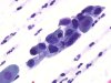
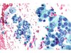
 | | Clusters and isolated parabasal cells with enlarged nuclei, single or multiple, with irregular nuclear outlines and coarse chromatin. HSIL. (obj. 20x) |
 | | Some basal cells, isolated or grouped, with enlarged nuclei, irregular nuclear outlines and coarse chromatin. HSIL. (obj. 20x) |
 | | Isolated atypical parabasal cells: ASC-H in this field but final diagnosis: HSIL. (obj. 20x) |
 | | HSIL: inflammatory smear with clusters of parabasal cells with enlarged, hyperchromatic, irregularly outlined nuclei. An immature metaplasia is a possibility. Final diagnosis: HSIL. (A: obj. 10x, B: obj. 20x) |
 | | Inflammatory smear with clusters of parabasal cells with enlarged, hyperchromatic, irregularly outlined nuclei. In this field: ASC-H. Final diagnosis: HSIL. (A: obj. 10x, B: obj. 20x) |
 | | Basal cells with enlarged nuclei and irregular chromatin, in between normal superficial and intermediate cells. These occasional cells do not allow a definite diagnosis of HSIL. It will be advisable to give a diagnosis of ASC-H on this picture. (A and B: obj. 20x) |


