Image | Statistics | Caption |
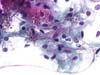
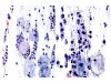
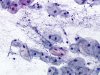
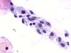
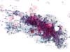 |  | Cervical and vaginal mycosis: many hyphae (arrows) and yeasts are visible at low magnification. (obj. 10x) |
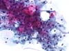 | | Cervical and vaginal mycosis: details of hyphae (arrows) and yeasts. (obj. 20x) |
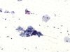
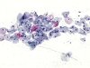
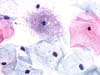
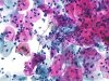 |  | Moniliasis (Candida albicans): hyphae and yeasts in an inflammatory background. (obj. 20x) |
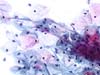 | | Moniliasis: details of hyphae and yeasts. (obj. 20x) |
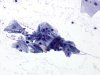
 | | Moniliasis: details of hyphae and yeasts. Compare sizes of yeasts and squamous cell nuclei. (obj. 40x) |
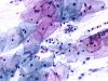
 | | Moniliasis: hyphae and yeasts may be detected at low magnification. (obj. 10x) |
 | | Moniliasis: hyphae and conidia are evident at medium magnification. (obj. 20x) |
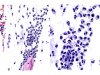
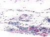 | | Ectocervix: fungal infection. Inflammatory background is present. (obj. 10x) |
 | | Ectocervix: many yeasts, pseudo-hyphae are less visible Inflammatory background is present. (A: obj. 20x, B: obj. 40x) |
 | | Moniliasis: hyphae and yeasts may be detected at low magnification. (obj. 10x) |
 | | Moniliasis: hyphae (purple arrow) and yeasts (ellipses) are obvious at this magnification. Intermediate cell with an enlarged nucleus (black arrow), suggesting an associated ASC-US. (obj. 20x) |
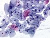
 | | Ectocervix: mycosis with yeasts, sometimes budding. (obj. 20x) |
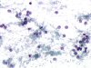
 | | Moniliasis: many spores with one visible filament, rare polymorphs and long bacilli. (obj. 20x) |
 | | Smear with marked cytolysis: yeasts, some polymorphs, naked nuclei and bacilli. (obj. 20x) |
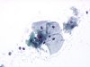 | | Smear with marked cytolysis: mycosis? Yeasts, squamous cells, naked nuclei and bacilli. (obj. 20x) |
 | | Ectocervix: pseudofilaments composed of mucus. An eosinophilic polymorph is present (circle). (A: obj. 20x, B: obj. 40x) |
 | | Mycosis: occasional yeasts (arrows): contamination during the sampling. Filamentous bacilli. (obj. 40x) |
 | | Ectocervix: normal flora with filamentous bacilli to be distinguished from pathogenic flora or from fungal filaments. (obj. 40x) |


