Image | Statistics | Caption |
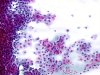
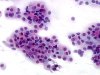
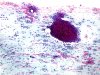
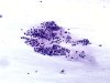
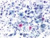
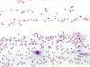
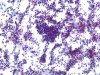
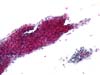
 |  | Menopausal atrophy with groups of basal cells and naked nuclei. (obj. 5x) |
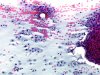
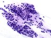
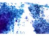
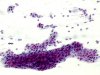 |  | Menopausal atrophic exocervical smear : sheets of basal cells and naked nuclei. (obj. 10x) |
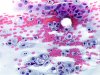
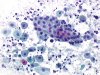
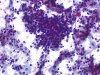
 | | Menopausal atrophy with groups of basal cells and naked nuclei. Sometimes it is difficult to separate these cells from atrophic endocervical cells. (obj. 10x) |
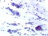
 | | Menopausal atrophy with large sheets of parabasal or basal cells and some naked nuclei. (obj. 10x) |
 | | Menopausal atrophy with large sheets of parabasal or basal cells and some naked nuclei. (obj. 20x) |
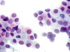 | | Menopausal atrophy with large sheets of parabasal or basal cells and some naked nuclei. (obj. 40x) |
 | | Menopausal atrophy with large sheets of parabasal or basal cells and some naked nuclei. (obj. 20x) |
 | | Menopausal atrophy with large sheets of parabasal or basal cells and some naked nuclei. (obj. 40x) |
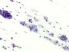
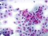
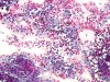
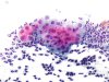
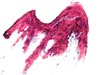
 | | Atrophic menopausal smear, bloody, with groups of basal squamous cells and numerous histiocytes. (obj. 5x) |
 | | Atrophic menopausal smear, bloody, with groups of basal squamous cells and numerous histiocytes (arrows). (obj. 10x) |
 | | Atrophic menopausal smear, bloody, with a group of basal squamous cells and many histiocytes. (obj. 20x) |
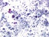
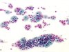
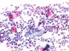
 | | Atrophic menopausal smear with rare basal squamous cells and numerous histiocytes, sometimes multinucleated. (obj. 10x) |
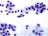
 | | Menopausal smear: columnar cells of classical morphology in the endocervical sample. (obj. 10x) |
 | | Menopausal smear: columnar cells of classical morphology in the endocervical sample. (obj. 20x) |
 | | Menopausal smear: columnar cells of classical morphology in the endocervical sample (honeycomb pattern). (obj. 40x) |
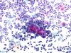
 | | Atrophic and inflammatory menopausal smear, containing small pseudo parakeratotic cells (red necrosis) and nuclear degenerative changes. One cell with an enlarged nucleus: ASC-H? Repeat after local estrogenic treatment. (obj. 20x) |
 | | Atrophic, inflammatory and necrotic menopausal smear, containing small pseudo parakeratotic cells (ellipse) and nuclear degenerative changes (arrows). Repeat after local estrogenic and anti-inflammatory treatment. (obj. 10x) |
 | | Atrophic, inflammatory and necrotic menopausal smear, containing small pseudo parakeratotic cells (red necrosis) and nuclear degenerative changes. Repeat after local estrogenic and anti-inflammatory treatment. (obj. 10x) |
 | | Atrophic, inflammatory and necrotic menopausal smear, containing small pseudo parakeratotic cells (red necrosis) and nuclear degenerative changes. A cluster of basal squamous or columnar cells. (obj. 10x) |
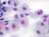
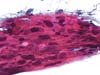
 | | Menopausal atrophy: large sheets of parabasal or deep intermediate cells. Atrophic epithelium is fragile explaining the bloody background. (obj. 10x) |
 | | Atrophic menopausal smear, showing no degenerative changes. (obj. 10x) |
 | | Bloody menopausal atrophic smear: some squamous parabasal cells, one with an enlarged nucleus: atrophic changes. To be checked after local oestrogen treatment. (obj. 10x) |
 | | Very inflammatory menopausal atrophic smear: a cluster of parabasal squamous cells without atypia. (obj. 20x) |
 | | Very inflammatory menopausal atrophic smear: a cluster of parabasal squamous cells and isolated cells with some nuclear atypia (slight enlargement) and cytoplasmic changes (acidophilia, vacuoles). Air-dried artefacts. To be check after local estrogenic treatment. (obj. 20x) |
 | | Very inflammatory menopausal atrophic smear: a cluster of parabasal squamous cells and isolated cells with some nuclear atypia (slight enlargement and hyperchromasia) and cytoplasmic changes (acidophilia, vacuoles). To be check after local estrogenic treatment. (obj. 20x) |
 | | Very inflammatory menopausal atrophic smear: a cluster of parabasal squamous cells and isolated cells with some nuclear atypia (slight increase in size and density) and cytoplasmic changes (acidophilia, vacuoles, cytophagia), to be distinguished from endometrial cells. (obj. 20x) |
 | | Atrophic, inflammatory and bloody menopausal smear, containing parabasal squamous cells and a thick cell aggregate, difficult to analyze: endometrial cells? Repeat after local estrogenic and anti-inflammatory treatment. (obj. 10x) |
 | | Atrophic, inflammatory and bloody menopausal smear, containing parabasal squamous cells and a thick cell aggregate, difficult to analyze: endometrial cells? Repeat after local estrogenic and anti-inflammatory treatment. (obj. 20x) |
 | | Menopausal atrophy with a large sheet of parabasal cells with round nuclei, containing clear chromatin and a distinct nucleolus. (obj. 20x) |
 | | Menopausal atrophy with large sheets of parabasal cells with round or oval nuclei, sometimes enlarged, in an inflammatory background. (obj. 20x, 2 different fields A and B) |
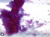
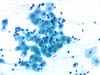
 | | Menopausal atrophic exocervical smear: sheets of basal cells with orangeophilic cytoplasm and rare naked nuclei. (obj. 10x) |
 | | Menopausal atrophic exocervical smear: sheets of basal cells with orangeophilic cytoplasm. (obj. 20x) |
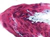
 | | Atrophic ectocervix (menopause): large sheet of squamous epithelium with irregular outlines. Orangeophilia and nuclear abnormalities to be reevaluated after oestrogenic treatment. Artefact preparation (air-dried) and delayed fixation. (obj. 10x) |
 | | Atrophic ectocervix (menopause): large sheet of squamous epithelium with irregular outlines. Orangeophilia and nuclear abnormalities to be reevaluated after local oestrogenic treatment. Artefact preparation (air-dried) and delayed fixation. (obj. 10x) |
 | | Atrophic ectocervix (menopause): large sheet of squamous epithelium with irregular outlines. Orangeophilia and nuclear abnormalities to be reevaluated after local oestrogenic treatment. Cellular details. Artefact preparation (air-dried) and delayed fixation. (obj. 20x) |
 | | Atrophic ectocervix (menopause): large sheet of squamous epithelium with irregular outlines. Orangeophilia and nuclear abnormalities to be reevaluated after local oestrogenic treatment. Cellular details. Artefact preparation (air-dried) and delayed fixation. (obj. 20x) |


