Image | Statistics | Caption |
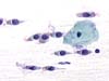
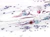 |  | Normal glandular cells either of the ciliated (arrows) or secretory type (ellipses). (obj. 10x) |
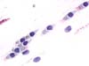
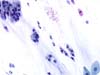 |  | Normal endocervical cells either of the ciliated or secretory type. (obj. 20x) |
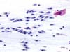
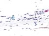 | | Normal endocervical cells of the ciliated type. (obj. 10x) |
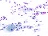
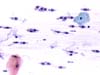 | | Normal endocervical cells of the ciliated type. (obj. 20x) |
 | | Normal glandular ciliated endocervical cells. Compare nuclear size and chromatin texture of ciliated cells and the squamous cell. (obj. 40x) |
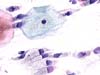
 | | Normal endocervical ciliated cells. Note basal plate (arrow) and cilia stained slightly pink. (obj. 40x) |
 | | Normal endocervical cells of the ciliated type. (obj. 20x) |
 | | Normal endocervical ciliated columnar cells with histiocytes (arrow). (obj. 20x) |
 | | Normal glandular endocervical cells either of the ciliated or secreting type (ellipse). Nuclei show quite the same morphology except for their outlines in secretory cells. (obj. 40x) |
 | | Mixture of normal glandular ciliated and secretory endocervical cells. (obj. 40x) |


