


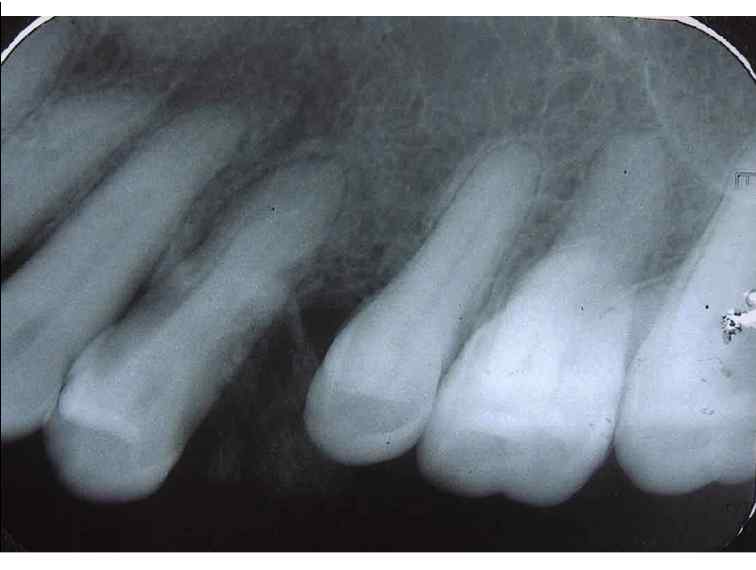
Figure 2: Intraoral periapical radiograph of the left maxillary premolar and molar region showing spacing between the premolars due to their displacement. Note the radiopacity of the soft tissues between the premolars.
25 avenue Tony Garnier CS 90627 69366, LYON CEDEX 07 France - Tel: +33 (0)4 72 73 84 85
© IARC 2025 - Terms of use - Privacy Policy.
© IARC 2025 - Terms of use - Privacy Policy.