


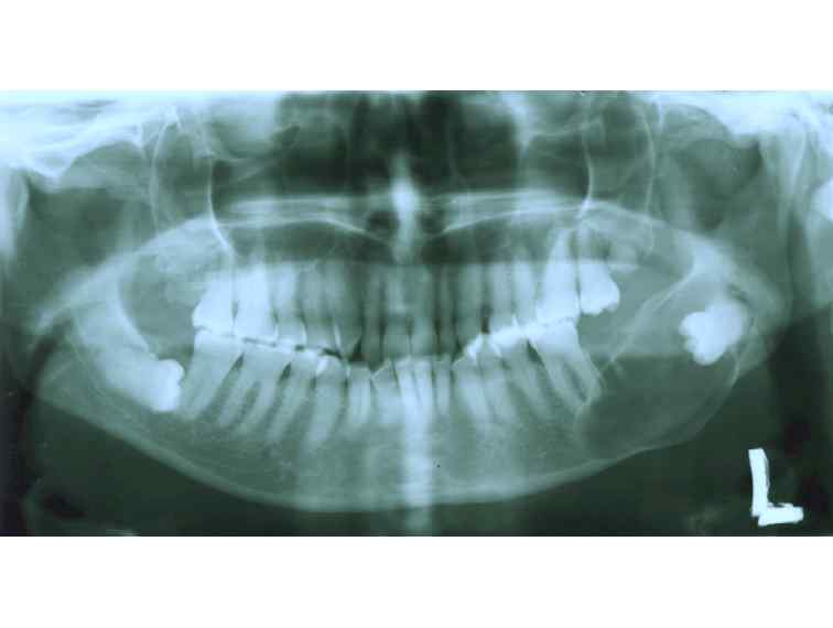
Figure 1: Dentigerous cyst: Panoramic radiograph showing a well-defined radiolucency with sclerotic border encompassing the crown of last molar on the left side of the lower jaw and extending from the mid part of ramus to the periapical region of the first molar. The second molar is missing. The lower border of mandible is expanded. The right mandiblar last molar is also horizontally impacted.
25 avenue Tony Garnier CS 90627 69366, LYON CEDEX 07 France - Tel: +33 (0)4 72 73 84 85
© IARC 2024 - Terms of use - Privacy Policy.
© IARC 2024 - Terms of use - Privacy Policy.