


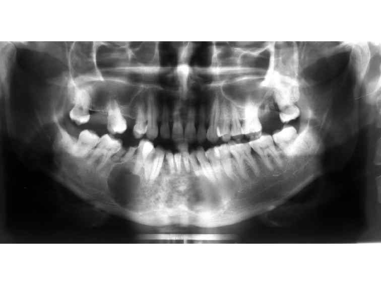
Figure 2: Ameloblastoma: Panoramic view shows a well-defined radiolucent lesion with scalloped margins extending from the distal aspect of the second mandibular premolar on the left side to the root of right mandibular first molar. Smaller loculation can be seen towards the centre with large loculation towards the periphery.
25 avenue Tony Garnier CS 90627 69366, LYON CEDEX 07 France - Tel: +33 (0)4 72 73 84 85
© IARC 2025 - Terms of use - Privacy Policy.
© IARC 2025 - Terms of use - Privacy Policy.