


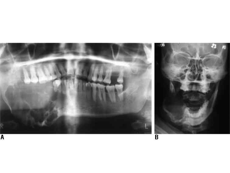
Figures 1: A: Pantomograph of a patient with ameloblastoma. Note the expansile multi locular radiolucency (soap bubble appearance) of the right side of mandible from the apex of the lateral incisor extending posteriorily to involve the ramus up to the condyle. B: Postero-anterior view of the face of the same patient, showing outward, downward and lingual expansion of the mandible.
25 avenue Tony Garnier CS 90627 69366, LYON CEDEX 07 France - Tel: +33 (0)4 72 73 84 85
© IARC 2025 - Terms of use - Privacy Policy.
© IARC 2025 - Terms of use - Privacy Policy.