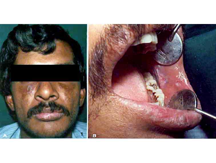
Figures 1: A: Extraoral photograph of a patient with discoid lupus erythematosus. Note the butterfly-shaped rash on the malar area. B: Intraoral photograph of the same patient showing erosive lesions surrounded by radiating white striae on the left buccal mucosa.
25 avenue Tony Garnier CS 90627 69366, LYON CEDEX 07 France - Tel: +33 (0)4 72 73 84 85
© IARC 2025 - Terms of use - Privacy Policy.
© IARC 2025 - Terms of use - Privacy Policy.


