


Home / Training / Manuals / Histopathology of the uterine cervix - digital atlas / Hysterectomy specimen - Gross examination study / video
Histopathology of the uterine cervix - digital atlas
Hysterectomy specimen - Gross examination study / video
Filter by language: English / Français / Portugues / 中文| Image/Video | Legend |
 | Gross morphology - a radical hysterectomy specimen, anterior side. The blue color is due to the injection of a dye needed for sentinel node identification. |
| Longitudinal section of the uterus (cutter knife is advised), Two hemi uterus are obtained. | |
| The vaginal section of the right hemi uterus is obtained. | |
| The vaginal section of the left hemi uterus is obtained. | |
| A gross section in the sagittal plane is performed. | |
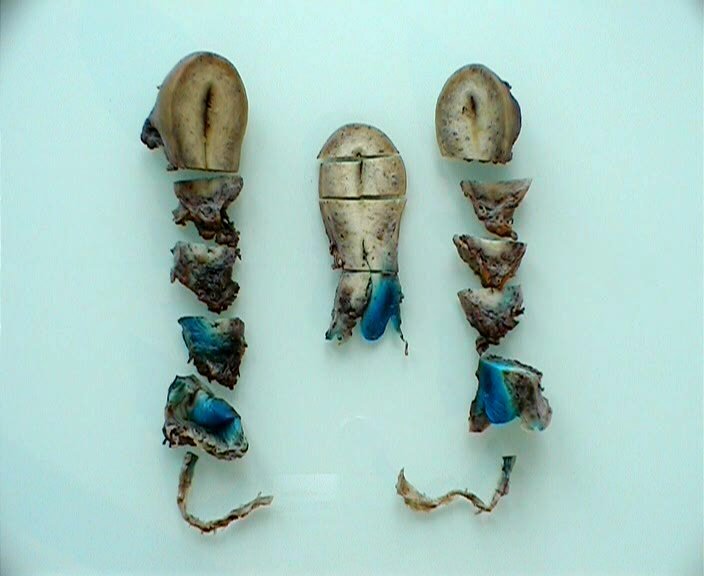
| Parallel sections in the transversal plane involving the cervical mass and the parametrium of each hemi uterus are obtained. | |
| Parallel sections in the transversal plane involving the cervical mass and the parametrium of each hemi uterus are obtained. | |
| Sampling of the gross section in the sagittal plane is performed: the cervical mass, the uterus wall, the use of big cassettes (70 x 50 x 15 mm) is sometimes required. | |
 | We obtain a full sample of the specimen of radical hysterectomy: the following are systematically included: the cervical mass, right parametrium, left parametrium, the uterus wall. |
25 avenue Tony Garnier CS 90627 69366, LYON CEDEX 07 France - Tel: +33 (0)4 72 73 84 85
© IARC 2026 - Terms of use - Privacy Policy.
© IARC 2026 - Terms of use - Privacy Policy.