


Home / Training / Manuals / Histopathology of the uterine cervix - digital atlas / Glossary Definitions
Histopathology of the uterine cervix - digital atlas
Glossary Definitions
Filter by language: English / Français / Portugues / 中文|
|
 |
|
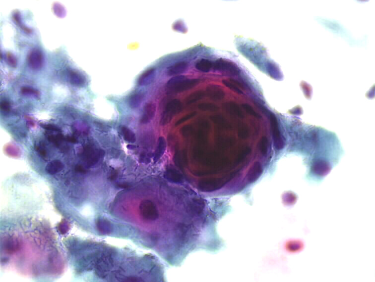
Cell-in-cell image These cell-in-cell figures are observed when squamous cells form swirls, like in onion bulbs. Cells in the middle are frequently keratinized. They may be seen in smears from non-neoplastic squamous epithelium (benign epithelial pearls) but more frequently in well differentiated and keratinised squamous cell carcinoma and some intraepithelial lesions (malignant squamous pearls). Cytopathology atlas |
25 avenue Tony Garnier CS 90627 69366, LYON CEDEX 07 France - Tel: +33 (0)4 72 73 84 85
© IARC 2024 - Terms of use - Privacy Policy.
© IARC 2024 - Terms of use - Privacy Policy.