Image | Statistics | Caption |
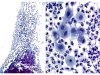
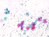 |  | Midly inflammatory smear: one binucleated cell (purple arrow) and small clear perinuclear halos (black arrows). Look for Trichomonas. (obj. 10x) |
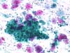
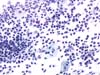 |  | Smear with marked inflammation: many polymorphs and histiocytes. (obj. 20x) |
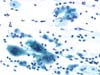 | | Multinucleated cells (histiocytes), must be distinguinshed from herpetic cells. (obj. 20x) |
 | | Multinucleated cell (histiocytes) more frequently seen in menopausal smears. (obj. 20x) |
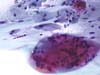
 | | Atrophic and inflammatory menopausal smear with isolated parabasal squamous cells. Three of them contain non specific cytoplasmic inclusions (previously reported as chlamydial infection). (field A: obj. 10x, field B: obj. 40x) |
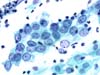
 | | Ectocervix: inflammatory background, sheet of metaplastic/repair type cells. (obj. 10x) |
 | | Inflammatory and bloody exocervical smear: sheet of repair cells with a clear nuclear chromatin and a visible nucleolus. (obj. 20x) |
 | | Repair cells (clear chromatin and nucleoli)? Immature or transitional metaplasia (some nuclear grooves)? (obj. 40x) |


