Image | Statistics | Caption |
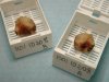
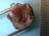 |  | Uterine cone, gross morphology. To identify the orientation of the specimen Indian ink of different colours may be use. |
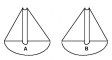 | | Scheme of conization sampling: sagittal median cut of the uterine cervix in two hemi-cones after fixation in 10% formaldehyde (day 2 to 4). Both hemi-cones are embedded in paraffin completely (day 4). |
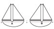 | | Scheme of conization sampling (day 5): Semi-serial sections are taken until the cervical canal disappears completely on the section. One section in every 10 to 30 cuts is kept and stained, the interval between two sections is around 100 to 200 microns. |
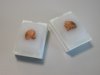
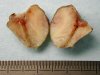 | | Uterine cervix, two hemi-cones after fixation in 10% buffered formaldehyde saline - 48 hours. |
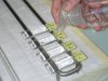
 | | Two hemi-cones put in cassettes (large cassettes 40x26x12mm). |
 | | Paraffin embedding of the two hemi-cones. |
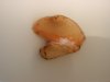 | | View of the block after paraffin embedding. |
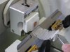
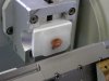 | | Installation of the block on the microtome (disposable blade). |
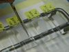
 | | The block is cut in semi-serial sections until the disappearance of the cervical canal - 5 microns thickness - note the ribbon obtained. |
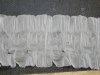 | | The ribbon. |
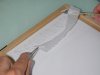 | | Setting the whole ribbon on a board. |
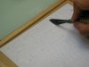
 | | Numbering the sections in chronological order. |
 | | Selection of one good quality section in every ten to twenty sections. Only these sections will be stained and mounted. The remaining of ribbons will be conserved until the final interpretation of the slides. |
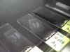
 | | Preparation of slides before placing sections. The glass slides (preferably pretreated by triethoxysilane) are covered with distilled water. |
 | | Placing the sections on the slides. |
 | | Spreading the sections on a hot plate (50°C) - removal of excess water. |


