Home / Training / Manuals / Colposcopy and treatment of cervical intraepithelial neoplasia: a beginners’ manual / Chapter 4: An introduction to colposcopy: indications for colposcopy, instrumentation, principles and documentation of results
 table 4.1: Indications for colposc...
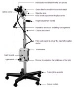 figure 4.1: Colposcope
...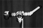 figure 4.2: Colposcope with a phot...
 figure 4.3: Colposcopy instrument ...
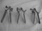 figure 4.4: Collins bivalve specul...
 figure 4.5: Vaginal side-wall retr...
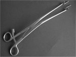 figure 4.6: Endocervical speculum<...
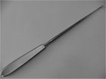 figure 4.7: Endocervical curette
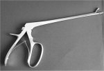 figure 4.8: Cervical punch biopsy ...
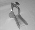 figure 4.9: Vaginal speculum cover...
 table 4.2: Preneoplastic intraepit...
Colposcopy and treatment of cervical intraepithelial neoplasia: a beginners’ manual, Edited by J.W. Sellors and R. Sankaranarayanan
Chapter 4: An introduction to colposcopy: indications for colposcopy, instrumentation, principles and documentation of results
Filter by language: English / Français / Español / Portugues / 中文- A colposcope is a low-power, stereoscopic, binocular field microscope with a powerful light source used for magnified visual examination of the uterine cervix to help in the diagnosis of cervical neoplasia.
- The most common indication of referral for colposcopy is positive screening tests (e.g., positive cytology, positive on visual inspection with acetic acid (VIA) etc.).
- The key ingredients of colposcopic examination are the observation of features of the cervical epithelium after application of normal saline, 3-5% dilute acetic acid, and Lugol’s iodine solution in successive steps.
- The characteristics of acetowhite changes, if any, on the cervix following the application of dilute acetic acid are useful in colposcopic interpretation and in directing biopsies.
- The colour changes in the cervix, following the application of Lugol’s iodine solution, depends on the presence or absence of glycogen in the epithelial cells. Areas containing glycogen turn brown or black; areas lacking glycogen remain colourless or pale or turn mustard or saffron yellow.
- It is important to carefully document the findings of colposcopic examination, immediately after the procedure, in a colposcopic record.
This chapter describes the indications for carrying out colposcopic examination of women, the instrumentation used for colposcopy, the basis of different colposcopic investigations and the methods of documentation of colposcopic findings. The step-by-step procedure of doing the colposcopic examination is described in the next chapter.
Indications for colposcopy
Given the availability of a colposcope and a trained colposcopist, there are a number of indications for this examination, of which positive cervical screening tests constitute the most frequent indication for colposcopy. The most common reason for referral of women for colposcopy is abnormal cervical cytology, usually discovered as a result of cytological screening (Table 4.1). Cytologically reported high-grade abnormalities such as high-grade cervical intraepithelial neoplasia (CIN 2 and CIN 3) may be associated with an underlying invasive squamous cell cervical cancer or adenocarcinoma. It is important that all women with high-grade abnormalities be referred immediately for diagnostic colposcopy. However, there is considerable variation in the management of women with low-grade abnormalities such as low-grade cervical intraepithelial neoplasia (CIN 1).
The referral criteria in some centres, for example in developing countries where colposcopy is available, allow for the immediate referral of women with low-grade abnormalities, whereas in other regions, for example in developed countries, they are called back for repeat cytology smears every six months for up to two years and only those with persistent or progressive abnormalities are referred. It should be emphasized that women with low-grade lesions (CIN 1) on their cytology smears have a higher probability of having a high-grade lesion that would be found at colposcopy; perhaps 15% for those with atypia and 20% with CIN 1 on cytology may harbour higher-grade lesions ( Shafi et al., 1997). It is advisable that women with any grade of CIN on cytology be referred for colposcopy in developing countries, in view of the possibility of reporting misclassification associated with cytology and poor compliance with follow-up.
Abnormal cytology results are tend to be quite worrying for a woman, as is a visit for a colposcopic examination. A few clinical caveats are worth mentioning. If the clinician observes characteristics of a cervix that looks suspicious, no matter what the cytology shows, it is advisable to refer the woman for colposcopic examination. Likewise, observation of an area of leukoplakia (hyperkeratosis) on the cervix should prompt a colposcopic examination, since the leukoplakia can not only obscure a lesion, but also preclude adequate cytological sampling of the area. It is still uncertain whether women with external anogenital warts are at increased risk of CIN; although it is clear that they should have routine cytology smears, it is not certain whether they would benefit from colposcopic examination ( Howard et al., 2001).
The potential roles of 3-5% acetic acid application and subsequent visual inspection of the cervix with magnification (VIAM) or without (VIA) and of visual inspection with Lugol’s iodine (VILI) are still under study as screening techniques ( University of Zimbabwe, JHPIEGO study, 1998; Denny et al., 2000; Belinson et al., 2001; Sankaranarayanan et al., 2001). Women who are positive for these tests may be referred for colposcopy to rule out underlying high-grade CIN and invasive cancer.
 table 4.1: Indications for colposc...
Instrumentation
Hinselmann (19.5) first described the basic colposcopic equipment and its use, establishing the foundation for the practice of colposcopy. A colposcope is a low-power, stereoscopic, binocular, field microscope with a powerful variable-intensity light source that illuminates the area being examined (Figure 4.1).
The head of the colposcope, also called the ‘optics carrier’, contains the objective lens (at the end of the head positioned nearest to the woman being examined), two ocular lenses or eyepieces (used by the colposcopist to view the cervix), a light source, green and/or blue filters to be interposed between the light source and the objective lens, a knob to introduce the filter, a knob to change the magnification of the objective lens, if the colposcope has multiple magnification facility and a fine focusing handle. The filter is used to remove red light, to facilitate the visualization of blood vessels by making them appear dark. Using a knob, the head of the colposcope can be tilted up and down to facilitate examination of the cervix. The distance between the two ocular lenses can be adjusted to suit the interpupillary distance of the provider, to achieve stereoscopic vision. Each ocular lens has dioptre scales engraved on it to facilitate visual correction of individual colposcopists. The height of the head from the floor can be adjusted by using the height adjustment knob, so that colposcopy can be carried out with the colposcopist comfortably seated, without strain to the back.
Modern colposcopes usually permit adjustable magnification, commonly 6x to 40x usually in steps such as, for example, 9x, 15x, 22x. Some sophisticated and expensive equipment may have electrical zoom capability to alter the magnification. Most simple colposcopes have a single fixed magnification level such as 6x, 9x, 10x, 12x or 15x. Most of the work with a colposcope can be accomplished within the magnification range of 6x to 15x. Lower magnification yields a wider view and greater depth of field for examination of the cervix. More magnification is not necessarily better, since there are certain trade-offs as magnification increases: the field of view becomes smaller, the depth of focus dimishes, and the illumination requirement increases. However, higher magnifications may reveal finer features such as abnormal blood vessels.
The location of the light bulb in the colposcope should be easily accessible to facilitate changing them when necessary. Some colposcopes have bulbs mounted in the head of the instrument; in others, these are mounted elsewhere and the light is delivered via a fibre-optic cable to the head of the colposcope. The latter arrangement can use brighter bulbs, but less overall illumination may result if the cables are bent or twisted. A colposcope may be fitted with halogen, xenon, tungsten or incandescent bulbs. Halogen bulbs are usually preferred, as they produce strong white light. The intensity of the light source may be adjusted with a knob.
Focusing the colposcope is accomplished by adjusting the distance between the objective lens and the woman by positioning the instrument at the right working distance. Colposcopes usually have fine focus adjustments so that, if the distance between the base of the scope and the woman is kept fixed, the focus of the scope may be altered slightly using the fine focusing handle. The working distance (focal length) between the objective lens and the patient is quite important - if it is too long (greater than 300 mm) it is hard for the colposcopist’s arms to reach the woman, and if it is too short (less than 200 mm), it may be difficult to use instruments like biopsy forceps while visualizing the target with the scope. A focal distance of 250 to 300 mm is usually adequate. Changing the power of the objective lenses alters the magnification and working distance.
Colposcopes are quite heavy and are either mounted on floor pedestals with wheels, suspended from a fixed ceiling mount, or fixed to the examination table or to a wall, sometimes with a floating arm to allow for easier adjustment of position. In developing countries, it is preferable to use colposcopes mounted vertically on a floor pedestal with wheels, as they are easier to handle and can be moved within or between clinics.
Accessories such as a monocular teaching side tube, photographic camera (Figure 4.2) and CCD video camera may be added to some colposcopes. However, these substantially increase the cost of the equipment. These accessories are added using a beam splitter in most colposcopes. The beam splitter splits the light beam in half and sends the same image to the viewing port and to the accessory port. Colpophotographic systems are useful for documentation of colposcopic findings and quality control. Teaching side tubes and videocolposcopy may be useful for real-time teaching and discussion of findings. With a modern CCD camera attached to a digitalizing port, it is possible to create high-resolution digital images of the colposcopic images.
 figure 4.1: Colposcope
...
 figure 4.2: Colposcope with a phot...
Examination table
The examination table allows the woman to be placed in a modified lithotomy position. The woman’s feet may be placed either in heel rests or the legs may be supported in knee crutches. Tables or chairs that can be moved up or down mechanically or electrically are more expensive and are not absolutely necessary either for colposcopic examination or to carry out treatment procedures guided by colposcopy.
Colposcopic instruments
The instruments needed for colposcopy are few and should be placed on an instrument trolley or tray (Figure 4.3) beside the examination table. The instruments required are: bivalve specula (Figure 4.4), vaginal side-wall retractor (Figure 4.5), cotton swabs, sponge-holding forceps, long (at least 20cm long) anatomical dissection forceps, endocervical speculum (Figure 4.6), endocervical curette (Figure 4.7), biopsy forceps (Figure 4.8), cervical polyp forceps and single-toothed tenaculum. In addition, the instrument tray may contain instruments necessary for treatment of CIN with cryotherapy or loop electrosurgical excision procedure (LEEP) (see chapters 11 and 12). The tray should also contain the consumables used for colposcopy and treatment.
In view of the different sizes of vagina, varying widths of bivalve specula should be available. One may use Cusco’s, Grave’s, Collin’s or Pedersen’s specula. One should use the widest possible speculum that can comfortably be inserted into the vagina to have optimal visualization of the cervix. Vaginal side-wall retractors are useful to prevent the lateral walls of a lax vagina from obstructing the view of the cervix. However, they may cause discomfort to the patient. An alternative approach is to use a latex condom on the speculum, the tip of which is opened with scissors 1 cm from the “nipple”(Figure 4.9). Sponge-holding forceps or long dissection forceps may be used to hold dry or moist cotton balls. The endocervical speculum or the long dissection forceps may be used to inspect the endocervical canal. The endocervical curette is used to obtain tissue specimens from the endocervix. Several types of sharp cervical biopsy punch forceps with long shafts (20-25 cm) such as Tischler-Morgan, Townsend or Kevrokian, are available. A single-toothed tenaculum or skin (iris) hook may be used to fix the cervix when obtaining a punch biopsy. Cervical polyps may be avulsed using the polyp forceps.
In view of the different sizes of vagina, varying widths of bivalve specula should be available. One may use Cusco’s, Grave’s, Collin’s or Pedersen’s specula. One should use the widest possible speculum that can comfortably be inserted into the vagina to have optimal visualization of the cervix. Vaginal side-wall retractors are useful to prevent the lateral walls of a lax vagina from obstructing the view of the cervix. However, they may cause discomfort to the patient. An alternative approach is to use a latex condom on the speculum, the tip of which is opened with scissors 1 cm from the “nipple”(Figure 4.9). Sponge-holding forceps or long dissection forceps may be used to hold dry or moist cotton balls. The endocervical speculum or the long dissection forceps may be used to inspect the endocervical canal. The endocervical curette is used to obtain tissue specimens from the endocervix. Several types of sharp cervical biopsy punch forceps with long shafts (20-25 cm) such as Tischler-Morgan, Townsend or Kevrokian, are available. A single-toothed tenaculum or skin (iris) hook may be used to fix the cervix when obtaining a punch biopsy. Cervical polyps may be avulsed using the polyp forceps.
 figure 4.3: Colposcopy instrument ...
 figure 4.4: Collins bivalve specul...
 figure 4.5: Vaginal side-wall retr...
 figure 4.6: Endocervical speculum<...
 figure 4.7: Endocervical curette
 figure 4.8: Cervical punch biopsy ...
 figure 4.9: Vaginal speculum cover...
Principles of colposcopy examination procedures
Saline technique
The key ingredients of colposcopic practice are the examination of the features of the cervical epithelium after application of saline, 3-5% dilute acetic acid and Lugol’s iodine solution in successive steps. The study of the vascular pattern of the cervix may prove difficult after application of acetic acid and iodine solutions. Hence the application of physiological saline before acetic acid and iodine application is useful in studying the subepithelial vascular architecture in great detail. It is advisable to use a green filter to see the vessels more clearly.
Principles of acetic acid test
The other key ingredient in colposcopic practice, 3-5% acetic acid, is usually applied with a cotton applicator (cotton balls held by sponge forceps, or large rectal or small swabs) or with a small sprayer. It helps in coagulating and clearing the mucus. Acetic acid is thought to cause swelling of the epithelial tissue, columnar and any abnormal squamous epithelial areas in particular. It causes a reversible coagulation or precipitation of the nuclear proteins and cytokeratins. Thus, the effect of acetic acid depends upon the amount of nuclear proteins and cytokeratins present in the epithelium. When acetic acid is applied to normal squamous epithelium, little coagulation occurs in the superficial cell layer, as this is sparsely nucleated. Though the deeper cells contain more nuclear protein, the acetic acid may not penetrate sufficiently and, hence, the resulting precipitation is not sufficient to obliterate the colour of the underlying stroma. Areas of CIN undergo maximal coagulation due to their higher content of nuclear protein and prevent light from passing through the epithelium. As a result, the subepithelial vessel pattern is obliterated and less easy to see and the epithelium appears white. This reaction is termed acetowhitening, and produces a noticeable effect compared with the normal pinkish colour of the surrounding normal squamous epithelium of the cervix, an effect that is commonly visible to the naked eye.
With low-grade CIN, the acetic acid must penetrate into the lower one-third of the epithelium (where most of the abnormal cells with high nuclear density are located). Hence, the appearance of the whiteness is delayed and less intense due to the smaller amount of nuclear protein compared to areas with high-grade CIN or preclinical invasive cancer. Areas of high-grade CIN and invasive cancer turn densely white and opaque immediately after application of acetic acid, due to their higher concentration of abnormal nuclear protein and the presence of large numbers of dysplastic cells in the superficial layers of the epithelium.
The acetowhite appearance is not unique to CIN and early cancer. It is also seen in other situations when increased nuclear protein is present: for example in immature squamous metaplasia, congenital transformation zone, in healing and regenerating epithelium (associated with inflammation), leukoplakia (hyperkeratosis) and condyloma. While the acetowhite epithelium associated with CIN and preclinical early invasive cancer is more dense, thick and opaque with well demarcated margins from the surrounding normal epithelium, the acetowhitening associated with immature squamous metaplasia and regenerating epithelium is less pale, thin, often translucent, and patchily distributed without well defined margins. Acetowhitening due to inflammation and healing is usually distributed widely in the cervix, not restricted to the transformation zone. The acetowhite changes associated with immature metaplasia and inflammatory changes quickly disappear, usually within 30-60 seconds.
Acetowhitening associated with CIN and invasive cancer quickly appears and persists for more than one minute. The acetic acid effect reverses much more slowly in high-grade CIN lesions and in early pre-clinical invasive cancer than in low-grade lesions, immature metaplasia and sub-clinical HPV changes. It may last for 2-4 minutes in the case of high-grade lesions and invasive cancer.
Acetowhitening also occurs in the vagina, external anogenital skin and anal mucosa (see Table 4.2). The acetowhite reaction varies in intensity, within and between patients. The reaction is often associated with other visual signs in the same area, and is not specific for intraepithelial preneoplasia. Invasive cancer may or may not be acetowhite; it usually has other distinguishing features that will alert the colposcopist. For these reasons, practical training is necessary to develop knowledge, skills and experience in colposcopy. Learning colposcopy requires more supervised practice than most other endoscopic procedures, because of the microscopic interpretation that must occur in vivo, in addition to the technical aspects of the endoscopic procedure.
As previously stated, the main goal of colposcopy is to detect the presence of high-grade CIN and invasive cancer. To effectively achieve this, the entire epithelium at risk should be well visualized, abnormalities should be identified accurately and assessed for their degree of abnormality, and appropriate biopsies must be taken. The colposcopic documentation and the biopsies taken by a colposcopist are important indicators for quality management in colposcopy clinics.
 table 4.2: Preneoplastic intraepit...
Principles of Schiller’s (Lugol’s) iodine test
The principle behind the iodine test is that original and newly formed mature squamous metaplastic epithelium is glycogenated, whereas CIN and invasive cancer contain little or no glycogen. Columnar epithelium does not contain glycogen. Immature squamous metaplastic epithelium usually lacks glycogen or, occasionally, may be partially glycogenated. Iodine is glycophilic and hence the application of iodine solution results in uptake of iodine in glycogen-containing epithelium. Therefore, the normal glycogen-containing squamous epithelium stains mahogany brown or black after application of iodine. Columnar epithelium does not take up iodine and remains unstained, but may look slightly discoloured due to a thin film of iodine solution; areas of immature squamous metaplastic epithelium may remain unstained with iodine or may be only partially stained. If there is shedding (or erosion) of superficial and intermediate cell layers associated with inflammatory conditions of the squamous epithelium, these areas do not stain with iodine and remain distinctly colourless in a surrounding black or brown background. Areas of CIN and invasive cancer do not take up iodine (as they lack glycogen) and appear as thick mustard yellow or saffron-coloured areas. Areas with leukoplakia (hyperkeratosis) do not stain with iodine. Condyloma may not, or occasionally may only partially, stain with iodine. We recommend the routine use of iodine application in colposcopic practice as this may help in identifying lesions overlooked during examination with saline and acetic acid and will help in delineating the anatomical extent of abnormal areas much more clearly, thereby facilitating treatment.
Documentation of colposcopic findings
The record of colposcopic findings for each visit should be documented carefully by the colposcopists themselves, immediately after the examination. This record, which can be stored on paper or electronically, forms the backbone of any medical record system that can be used for continuing patient care and performance. An example of a colposcopy record containing the important attributes of a colposcopic assessment is shown in Appendix 1. Colposcopists or clinics can adapt this form to suit their needs; the structured format is intended to prompt the colposcopist to use quantitative data, whenever possible, and to capture qualitative data in a drawing. Colposcopists, even in the same clinic, generally record their findings in a wide variety of ways. Various experts have recommended standardized representations of colposcopic findings in a drawing; the symbolic representations suggested by René Cartier are a good example of what may be useful in this context ( Cartier & Cartier, 1993).
Since an examination of the entire lower genital tract should be performed whenever a woman is referred for colposcopy, the colposcopist must be able to record the clinical findings in the vaginal, vulvar, perianal and anal epithelium. These findings can be combined with the cervical record on one page or kept on a separate page.


