


Home / Training / Manuals / A practical manual on visual screening for cervical neoplasia / Testing and reporting the results of visual inspection with 5% acetic acid (VIA)
 /
/ 
Instruments and materials required
Preparation of 5% dilute acetic acid
5% acetic acid is prepared by adding 5 ml of glacial acetic acid into 95 ml of distilled water.
If vinegar bought from a store is used, check the strength to ensure that it is 5%.
Test provider skills
The test provider must have a good knowledge of the anatomy, physiology and pathology of the cervix in relation to its visual examination. He/she should to know the clinical features of benign conditions, inflammation, precancerous lesions and invasive cancer of the cervix.
Procedure
Women coming for testing should have the screening procedure explained to them in detail. Written informed consent should be obtained before screening. An example of a written informed consent form is given in Appendix 2. Relevant obstetric and gynaecological history should be obtained and recorded with the help of a form for this purpose (Appendix 3). The woman should be reassured that the procedure is painless, and every effort should be made to ensure that she is fully relaxed and remains at ease during testing.
The woman is invited to lie down in a modified lithotomy position on a couch with leg rests or knee crutches or stirrups. After proper positioning of the woman, observe if there is any vaginal discharge. Observe the external genitalia, and perineal region for any signs of excoriations, oedema, vesicles, papules, sores, ulceration and warts. Look for any swelling in the inguinal/femoral region.
Afterwards, gently introduce a sterile vaginal speculum, which has been immersed in warm water and open the blades of the speculum to view the cervix. Adjust the light source so that there is adequate light in the vagina and on the cervix. As the speculum is gently opened and the lips are fixed, the cervix comes into view. Observe the size and shape of the cervix.
Identify the external os, columnar epithelium (red in colour), squamous epithelium (pink) and the squamocolumnar junction. Proceed to identify the transformation zone, the upper limit of which is formed by the squamocolumnar junction. Remember that cervical neoplasias occur in the transformation zone nearest to the squamocolumnar junction.
Look for ectropion, cervical polyp, nabothian cysts, healed laceration of the cervical lips, leukoplakia, condylomata and signs of cervicitis. You may note that in post menopausal women, the cervix appears pale and brittle, due to thinning and atrophy of the squamous epithelium. Assess the characteristics of discharge in terms of quantity, colour, odour and thickness. Thread-like, thin mucinous discharge from the external os indicates ovulation. If heavy blood flow through the external os is observed in women during menstruation, they may be subjected to VIA after 5-15 days.
In ectropion, the cervix has a large area of red appearance around the external os and the squamocolumnar junction is far away from the os. Nabothian cysts appear as bulging blue-white or yellow-white nodules, having a smooth delicate lining with branching blood vessels. In some women, nabothian cysts can become large and distort the shape of the cervix. A cervical polyp appears as a smooth mass protruding from the cervical canal beyond the external os, which may appear dark red or pink-white. Sometimes a necrotic polyp resembles a cervical cancer. Healed lacerations appear as tears on the lips of the cervix, with the external os appearing irregular. Leukoplakia appears as a smooth-surfaced, white area on the cervix that cannot be removed or scraped off. Cervical condylomata appear as raised, grey-white areas within or outside the transformation zone in the squamous epithelium and may be accompanied by similar lesions in the vagina and vulva.
Look for small blisters containing fluid or multiple, small ulcers on the cervix. Extensive erosive red areas may be present on the cervix, extending to the vagina in instances of severe cervical infection and inflammation. Observe whether there is any bleeding from the cervix, especially on touch, or ulceroproliferative growth. A very early invasive cancer may present as a rough, reddish, granular area, that may bleed on touch. More advanced invasive cancers may present as a large exophytic growth with an ulceroproliferative, bulging mass with polypoid or papillary excrescences, arising from the cervix or as a predominantly ulcerating growth replacing most of the cervix. In both of these types, bleeding on touch and necrosis are predominant clinical features. Foul-smelling discharge is also common due to superadded infection. Occasionally, invasive cancer can present as an infiltrating lesion with a grossly enlarged irregular cervix.
Now, gently, but firmly, apply 5% acetic acid using a cotton swab soaked in acetic acid. The secretions should be gently wiped off. The swabs after use should be disposed of in the waste bucket. The curdy-white discharge associated with candidiasis is particularly sticky, and if particular care is not taken to remove it properly, it may mimic an acetowhite lesion, thus leading to a false-positive result. After removing the swab, carefully look at the cervix to see whether any white lesions appear, particularly in the transformation zone close to the squamocolumnar junction, or dense, non-removable acetowhite areas in the columnar epithelium. The results one minute after application of acetic acid should be reported. Note how rapidly the acetowhite lesion appears and then disappears.
Carefully observe:
Conclusion of the examination
Contaminated swabs, gauze and other waste material should be disposed of in the plastic bag in a plastic bucket.
Withdraw the speculum gently, and inspect the vaginal walls for condyloma and acetowhite lesions. Before removing the soiled gloves, immerse the hands briefly in a container filled with 0.5% chlorine solution. Decontaminate the used gloves by soaking in the 0.5% chlorine in a plastic bucket for 10 minutes. Preparation of 0.5% chlorine solution is described in Appendix 4.
The speculum and other instruments used for VIA should be immersed in 0.5% chlorine solution for 10 minutes' decontamination, before cleaning with detergent and water. The cleaned instruments may be reused after high-level disinfection by immersing them in boiling water for 20 minutes or by sterilizing the instruments using an autoclave.
Documentation of findings and advising the woman
Carefully document the outcome of testing in the reporting form (Appendix 3). Explain the outcome of the test to the woman, as well as any further course of follow-up actions. If the test is negative, the woman is reassured and she may be advised to repeat testing after five years. If the test is positive, she should be referred for further investigations such as colposcopy and biopsy as well as treatment for any confirmed lesions. If invasive cancer is suspected, she should be referred to a cancer diagnosis and treatment facility.
Reporting the outcome of VIA
VIA negative (-)
VIA screening is reported as negative in the case of any of the following observations:
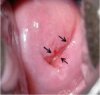
fig 2.1: VIA negative: No acetowhite area seen. Note the advancing edges of squamous metaplasia in the anterior and posterior lips (arrows).
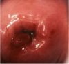
fig 2.2: VIA negative. There are no acetowhite areas on the polyp and the cervix after the application of acetic acid.
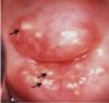
fig 2.3: VIA negative. The nabothian cysts appear as pimble- or button-like areas after the application of acetic acid.
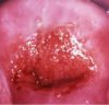
fig 2.4: VIA negative: There is dot-like acetowhitening in the columnar epithelium in the anterior lip. The squamocolumnar junction is fully visible.
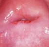
fig 2.5: VIA negative: There are ill-defined pinkish-white and cloudy-white areas with indefinite margins blending with the rest of the epithelium. The squamocolumnar junction is fully visible.
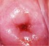
fig 2.6: VIA negative: There is an ill-defined pinkish-white hue with indefinite margins blending with the rest of the epithelium. The squamocolumnar junction is fully visible.
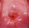
fig 2.7: VIA negative: There is an ill-defined pinkish-white hue, with indefinite margins, blending with the rest of the epithelium. The squamocolumnar junction is fully visible.
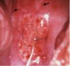
fig 2.8: VIA negative: There are pale white, satellite, geographical lesions with angular margins (narrow arrows) far away from the squamocolumnar junction (dense arrow). Note the streak-like acetowhitening in the columnar epithelium (within the oval area).
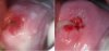
fig 2.9: VIA negative: There is dense, thick, mucus on the cervix before the application of acetic acid. After the application of acetic acid, the mucus is cleared and the squamocolumnar junction becomes prominent.
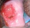
fig 2.10: VIA negative: The squamocolumnar junction is prominent after the application of acetic acid. Note the ectropion.
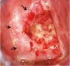
fig 2.11: VIA negative: The cervix is unhealthy, inflamed with ulceration, necrosis, bleeding and inflammatory exudate.There is ill-defined, diffuse, pinkish-white acetowhitening with indefinite margins blending with the rest of epithelium (arrows).
VIA positive (+)
The VIA test outcome is reported as positive in any of the following situations:
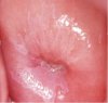
fig 2.12: VIA positive: There is a well-defined, opaque acetowhite area, with irregular digitating margins, in the anterior and posterior lips abutting the squamocolumnar junction and extending into the cervical canal.
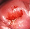
fig 2.13: VIA positive: There is a well-defined, opaque acetowhite area, with bleeding on touch, in the anterior lip, abutting the squamocolumnar junction, which is fully visible.
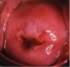
fig 2.14: VIA positive: There is a well-defined, opaque acetowhite area, with regular margins, in the anterior lip, abutting the squamocolumnar junction, which is fully visible.
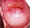
fig 2.15: VIA positive: There is a well-defined, opaque acetowhite area, with regular margins, in the lower lip, abutting the squamocolumnar junction, which is fully visible.
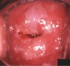
fig 2.16: VIA positive: There is a well-defined, opaque acetowhite area, with regular margins, in the anterior lip, abutting the squamocolumnar junction, which is fully visible. Note the satellite lesions in the lower lip.
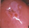
fig 2.17: VIA positive: There is a well-defined, opaque acetowhite area, with regular margins, in the anterior lip, abutting the squamocolumnar junction, which is fully visible. Note the somewhat ill-defined white area in the lower lip. The lesion is extending into the cervical canal.
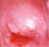
fig 2.18: VIA positive: There is a well-defined, dull, dense, opaque acetowhite area in the anterior lip abutting the squamocolumnar junction which is fully visible.
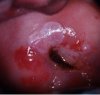
fig 2.19: VIA positive: There is well-defined, dull, dense, opaque acetowhite area, with raised and rolled-out margins in the anterior lip abutting the squamocolumnar junction which is fully visible. The lesion is extending into the cervical canal.
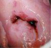
fig 2.20: VIA positive: There is a well-defined, dull, dense, opaque acetowhite area in the posterior lip extending into the endocervical canal.
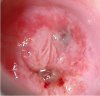
fig 2.21: VIA positive: There is an acetowhite area in the columnar epithelium in the anterior and posterior lips.
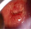
fig 2.22: VIA positive: There are dense acetowhite areas in the columnar epithelium in the anterior lip.
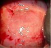
fig 2.23: VIA positive: There is a dense acetowhite area all over the cervix involving all the four quadrants and extending into the cervical canal.
VIA positive, invasive cancer
The test outcome is scored as invasive cancer when:
There is a clinically visible ulcero-proliferative growth on the cervix that turns densely white after application of acetic acid and bleeds on touch (Figure 2.24-2.27).
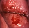
fig 2.24: VIA positive, invasive cancer: There is a dull, opaque, dense acetowhite area, with raised and rolled-out margins, irregular surface and bleeding on touch in the posterior lip. The lesion is extending into the cervical canal. The bleeding obliterates acetowhitening.
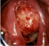
fig 2.25: VIA positive, invasive cancer: There is a proliferative growth with dense acetowhitening and bleeding.
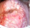
fig 2.26: VIA positive, invasive cancer: There is a dense acetowhite area with irregular surface contour.
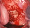
fig 2.27: VIA positive, invasive cancer: There is an ulceroproliferative growth with acetowhitening and bleeding.
Self-evaluation of the test providers
The test providers are encouraged to correlate the results of their VIA testing with those of colposcopy and histology. They are strongly advised to participate in the colposcopy sessions with doctors and review the findings. Such measures improve the skills of the test providers. A benchmark to assess one's own skill is to estimate what proportion of women examined are scored as aceto-positive and what proportion of aceto-positive women are ultimately diagnosed with CIN. A sufficiently skilled examiner will categorize 8-15% of women examined as aceto-positive, and 20-30% of the acetowhite lesions identified on VIA by the test provider harbour CIN of any grade.
A practical manual on visual screening for cervical neoplasia
Chapter 2 / Testing and reporting the results of visual inspection with 5% acetic acid (VIA)
Filter by language: English / Français / Español / Turkish / /
- Examination table with knee crutches or leg rests or stirrups;
- Good light source (preferably a bright halogen lamp that can be easily directed at the cervix or a bright halogen torch light);
- Sterile bivalved speculum: Cusco's, Grave's or Collin's;
- Pair of gloves;
- Cotton swabs, cotton-tipped buds, gauze;
- Ring forceps, pickup forceps;
- 5% freshly prepared acetic acid or vinegar (check the strength of acetic acid in vinegar);
- A steel/plastic container with 0.5% chlorine solution in which to immerse the gloves;
- A plastic bucket or container with 0.5% chlorine solution to decontaminate instruments;
- A plastic bucket with a polythene bag to dispose of contaminated swabs and other waste items.
If vinegar bought from a store is used, check the strength to ensure that it is 5%.
The woman is invited to lie down in a modified lithotomy position on a couch with leg rests or knee crutches or stirrups. After proper positioning of the woman, observe if there is any vaginal discharge. Observe the external genitalia, and perineal region for any signs of excoriations, oedema, vesicles, papules, sores, ulceration and warts. Look for any swelling in the inguinal/femoral region.
Afterwards, gently introduce a sterile vaginal speculum, which has been immersed in warm water and open the blades of the speculum to view the cervix. Adjust the light source so that there is adequate light in the vagina and on the cervix. As the speculum is gently opened and the lips are fixed, the cervix comes into view. Observe the size and shape of the cervix.
Identify the external os, columnar epithelium (red in colour), squamous epithelium (pink) and the squamocolumnar junction. Proceed to identify the transformation zone, the upper limit of which is formed by the squamocolumnar junction. Remember that cervical neoplasias occur in the transformation zone nearest to the squamocolumnar junction.
Look for ectropion, cervical polyp, nabothian cysts, healed laceration of the cervical lips, leukoplakia, condylomata and signs of cervicitis. You may note that in post menopausal women, the cervix appears pale and brittle, due to thinning and atrophy of the squamous epithelium. Assess the characteristics of discharge in terms of quantity, colour, odour and thickness. Thread-like, thin mucinous discharge from the external os indicates ovulation. If heavy blood flow through the external os is observed in women during menstruation, they may be subjected to VIA after 5-15 days.
In ectropion, the cervix has a large area of red appearance around the external os and the squamocolumnar junction is far away from the os. Nabothian cysts appear as bulging blue-white or yellow-white nodules, having a smooth delicate lining with branching blood vessels. In some women, nabothian cysts can become large and distort the shape of the cervix. A cervical polyp appears as a smooth mass protruding from the cervical canal beyond the external os, which may appear dark red or pink-white. Sometimes a necrotic polyp resembles a cervical cancer. Healed lacerations appear as tears on the lips of the cervix, with the external os appearing irregular. Leukoplakia appears as a smooth-surfaced, white area on the cervix that cannot be removed or scraped off. Cervical condylomata appear as raised, grey-white areas within or outside the transformation zone in the squamous epithelium and may be accompanied by similar lesions in the vagina and vulva.
Look for small blisters containing fluid or multiple, small ulcers on the cervix. Extensive erosive red areas may be present on the cervix, extending to the vagina in instances of severe cervical infection and inflammation. Observe whether there is any bleeding from the cervix, especially on touch, or ulceroproliferative growth. A very early invasive cancer may present as a rough, reddish, granular area, that may bleed on touch. More advanced invasive cancers may present as a large exophytic growth with an ulceroproliferative, bulging mass with polypoid or papillary excrescences, arising from the cervix or as a predominantly ulcerating growth replacing most of the cervix. In both of these types, bleeding on touch and necrosis are predominant clinical features. Foul-smelling discharge is also common due to superadded infection. Occasionally, invasive cancer can present as an infiltrating lesion with a grossly enlarged irregular cervix.
Now, gently, but firmly, apply 5% acetic acid using a cotton swab soaked in acetic acid. The secretions should be gently wiped off. The swabs after use should be disposed of in the waste bucket. The curdy-white discharge associated with candidiasis is particularly sticky, and if particular care is not taken to remove it properly, it may mimic an acetowhite lesion, thus leading to a false-positive result. After removing the swab, carefully look at the cervix to see whether any white lesions appear, particularly in the transformation zone close to the squamocolumnar junction, or dense, non-removable acetowhite areas in the columnar epithelium. The results one minute after application of acetic acid should be reported. Note how rapidly the acetowhite lesion appears and then disappears.
Carefully observe:
- The intensity of the white colour of the acetowhite lesion: if it is shiny-white, cloudy-white, pale-white, dull-white;
- The borders and demarcations of the white lesion: distinctly clear and sharp or indistinct diffuse margins; raised or flat margins; regular or irregular margins;
- Whether the lesions are uniformly white in colour, or the colour intensity varies across the lesion, or if there are areas of erosion within the lesion;
- Location of the lesion: is it in, near, or far away from the transformation zone? Is it abutting (touching) the squamocolumnar junction? Does it extend into the endocervical canal? Does it occupy the entire, or part of, the transformation zone? Does it involve the entire cervix (which usually indicates early preclinical invasive cancer)?
- Size (extent or dimensions) and number of the lesions.
Withdraw the speculum gently, and inspect the vaginal walls for condyloma and acetowhite lesions. Before removing the soiled gloves, immerse the hands briefly in a container filled with 0.5% chlorine solution. Decontaminate the used gloves by soaking in the 0.5% chlorine in a plastic bucket for 10 minutes. Preparation of 0.5% chlorine solution is described in Appendix 4.
The speculum and other instruments used for VIA should be immersed in 0.5% chlorine solution for 10 minutes' decontamination, before cleaning with detergent and water. The cleaned instruments may be reused after high-level disinfection by immersing them in boiling water for 20 minutes or by sterilizing the instruments using an autoclave.
- No acetowhite lesions are observed on the cervix (Figure 2.1).
- Polyps protrude from the cervix with bluish-white acetowhite areas (Figure 2.2).
- Nabothian cysts appear as button-like areas, as whitish acne, or pimples (Figure 2.3).
- Dot-like areas are present in the endocervix, which are due to grape-like columnar epithelium staining with acetic acid (Figure 2.4).
- There are shiny, pinkish-white, cloudy-white, bluish-white, faint patchy, or doubtful lesions with ill-defined, indefinite margins, blending with the rest of the cervix (Figures 2.5 - 2.7).
- Angular, irregular, digitating acetowhite lesions, resembling geographical regions, distant (detached) from the squamocolumnar junction (satellite lesions) (Figure 2.8).
- Faint line-like or ill-defined acetowhitening is seen at the squamocolumnar junction (Figures 2.8-2.10).
- Streak-like acetowhitening is visible in the columnar epithelium (Figure 2.8).
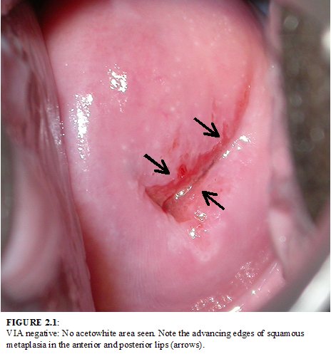
fig 2.1: VIA negative: No acetowhite area seen. Note the advancing edges of squamous metaplasia in the anterior and posterior lips (arrows).

fig 2.2: VIA negative. There are no acetowhite areas on the polyp and the cervix after the application of acetic acid.
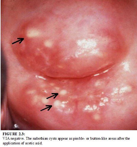
fig 2.3: VIA negative. The nabothian cysts appear as pimble- or button-like areas after the application of acetic acid.
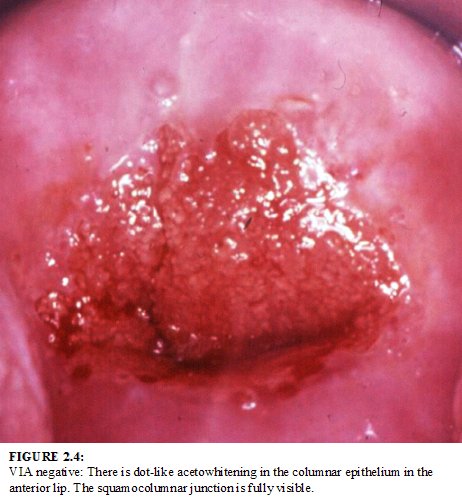
fig 2.4: VIA negative: There is dot-like acetowhitening in the columnar epithelium in the anterior lip. The squamocolumnar junction is fully visible.
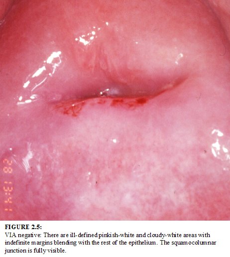
fig 2.5: VIA negative: There are ill-defined pinkish-white and cloudy-white areas with indefinite margins blending with the rest of the epithelium. The squamocolumnar junction is fully visible.
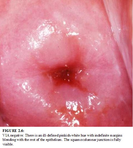
fig 2.6: VIA negative: There is an ill-defined pinkish-white hue with indefinite margins blending with the rest of the epithelium. The squamocolumnar junction is fully visible.
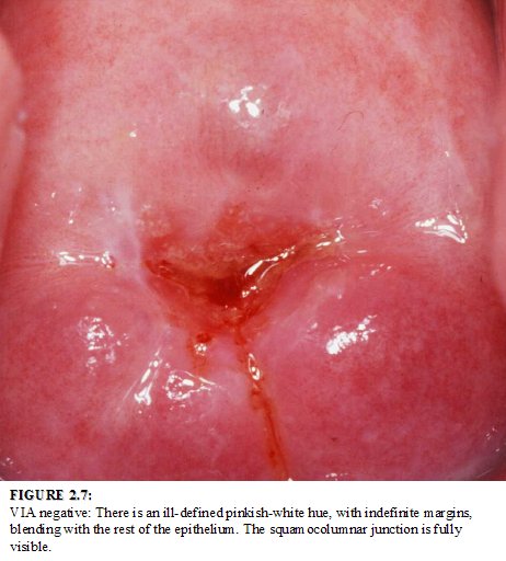
fig 2.7: VIA negative: There is an ill-defined pinkish-white hue, with indefinite margins, blending with the rest of the epithelium. The squamocolumnar junction is fully visible.
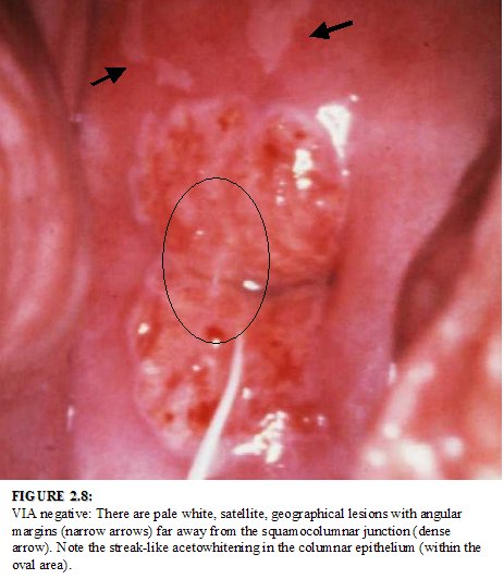
fig 2.8: VIA negative: There are pale white, satellite, geographical lesions with angular margins (narrow arrows) far away from the squamocolumnar junction (dense arrow). Note the streak-like acetowhitening in the columnar epithelium (within the oval area).
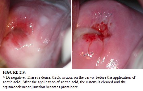
fig 2.9: VIA negative: There is dense, thick, mucus on the cervix before the application of acetic acid. After the application of acetic acid, the mucus is cleared and the squamocolumnar junction becomes prominent.
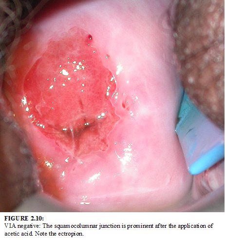
fig 2.10: VIA negative: The squamocolumnar junction is prominent after the application of acetic acid. Note the ectropion.

fig 2.11: VIA negative: The cervix is unhealthy, inflamed with ulceration, necrosis, bleeding and inflammatory exudate.There is ill-defined, diffuse, pinkish-white acetowhitening with indefinite margins blending with the rest of epithelium (arrows).
- There are distinct, well defined, dense (opaque, dull- or oyster-white) acetowhite areas with regular or irregular margins, close to or abutting the squamocolumnar junction in the transformation zone or close to the external os if the squamocolumnar junction is not visible (Figure 2.12- 2.20).
- Strikingly dense acetowhite areas are seen in the columnar epithelium (Figure 2.21-2.22).
- The entire cervix becomes densely white after the application of acetic acid (Figure 2.23)
- Condyloma and leukoplakia occurr close to the squamocolumnar junction, turning intensely white after application of acetic acid.
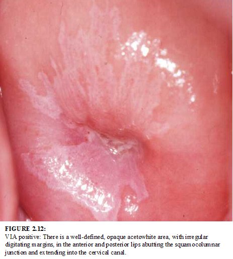
fig 2.12: VIA positive: There is a well-defined, opaque acetowhite area, with irregular digitating margins, in the anterior and posterior lips abutting the squamocolumnar junction and extending into the cervical canal.
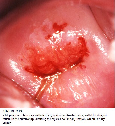
fig 2.13: VIA positive: There is a well-defined, opaque acetowhite area, with bleeding on touch, in the anterior lip, abutting the squamocolumnar junction, which is fully visible.
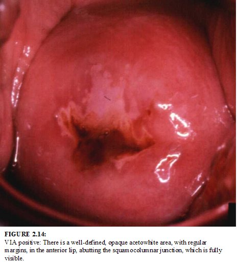
fig 2.14: VIA positive: There is a well-defined, opaque acetowhite area, with regular margins, in the anterior lip, abutting the squamocolumnar junction, which is fully visible.
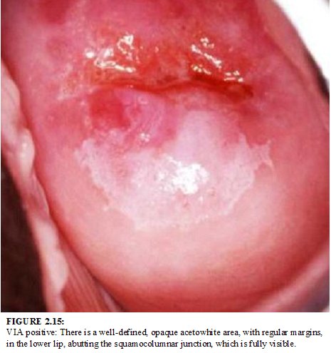
fig 2.15: VIA positive: There is a well-defined, opaque acetowhite area, with regular margins, in the lower lip, abutting the squamocolumnar junction, which is fully visible.
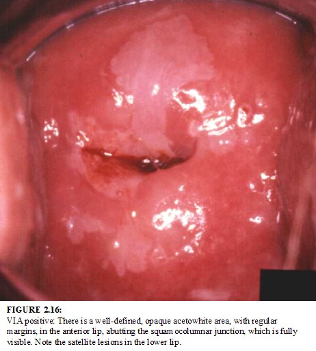
fig 2.16: VIA positive: There is a well-defined, opaque acetowhite area, with regular margins, in the anterior lip, abutting the squamocolumnar junction, which is fully visible. Note the satellite lesions in the lower lip.
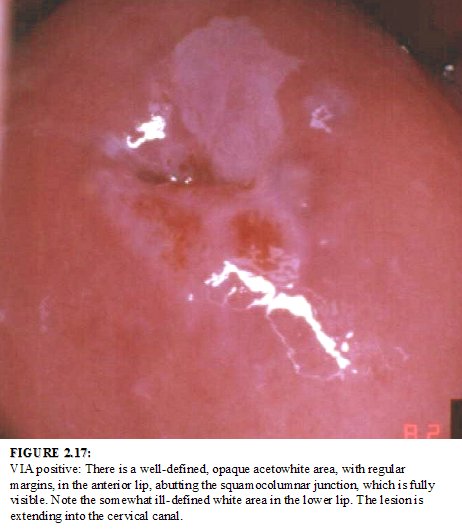
fig 2.17: VIA positive: There is a well-defined, opaque acetowhite area, with regular margins, in the anterior lip, abutting the squamocolumnar junction, which is fully visible. Note the somewhat ill-defined white area in the lower lip. The lesion is extending into the cervical canal.
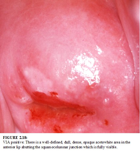
fig 2.18: VIA positive: There is a well-defined, dull, dense, opaque acetowhite area in the anterior lip abutting the squamocolumnar junction which is fully visible.
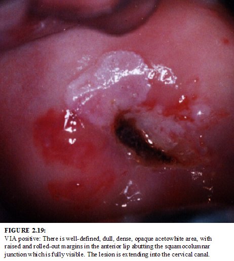
fig 2.19: VIA positive: There is well-defined, dull, dense, opaque acetowhite area, with raised and rolled-out margins in the anterior lip abutting the squamocolumnar junction which is fully visible. The lesion is extending into the cervical canal.

fig 2.20: VIA positive: There is a well-defined, dull, dense, opaque acetowhite area in the posterior lip extending into the endocervical canal.
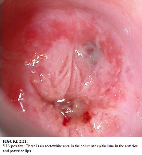
fig 2.21: VIA positive: There is an acetowhite area in the columnar epithelium in the anterior and posterior lips.
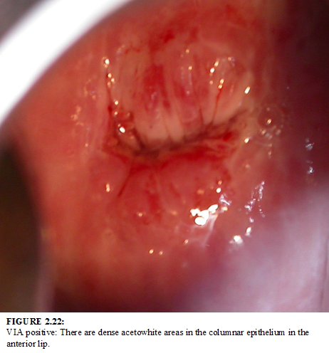
fig 2.22: VIA positive: There are dense acetowhite areas in the columnar epithelium in the anterior lip.
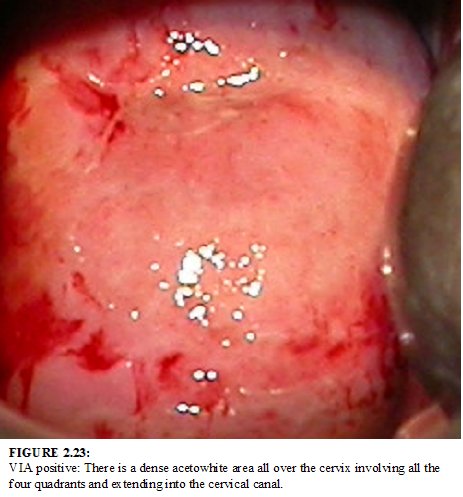
fig 2.23: VIA positive: There is a dense acetowhite area all over the cervix involving all the four quadrants and extending into the cervical canal.
There is a clinically visible ulcero-proliferative growth on the cervix that turns densely white after application of acetic acid and bleeds on touch (Figure 2.24-2.27).
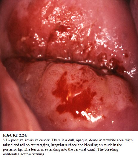
fig 2.24: VIA positive, invasive cancer: There is a dull, opaque, dense acetowhite area, with raised and rolled-out margins, irregular surface and bleeding on touch in the posterior lip. The lesion is extending into the cervical canal. The bleeding obliterates acetowhitening.
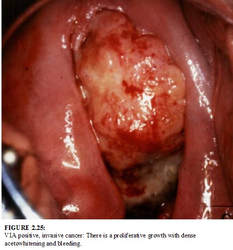
fig 2.25: VIA positive, invasive cancer: There is a proliferative growth with dense acetowhitening and bleeding.
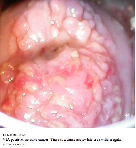
fig 2.26: VIA positive, invasive cancer: There is a dense acetowhite area with irregular surface contour.
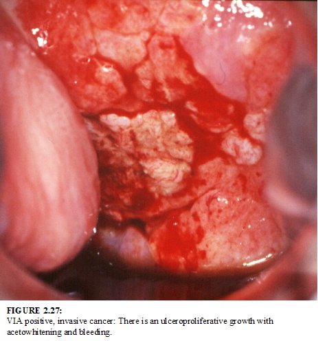
fig 2.27: VIA positive, invasive cancer: There is an ulceroproliferative growth with acetowhitening and bleeding.