


Learning colposcopy
Colposcopic appearance of normal cervix
The colposcopic examination
Detection of infections & benign conditions of cervix
Detection of cervical neoplasias
Treatment of cervical intraepithelial neoplasia
Treatment by cryotherapy
Treatment by thermal ablation
Treatment by LLETZ (LEEP)
Treatment by cold-knife conization (CKC)
Cases
Normal
Squamous metaplasia and ectropion
Inflammation and cervicitis
Low grade
High grade
Early and advanced cancers
Miscellaneous
Post treatment
Search with IFCPC criteria
Search with Swede score criteria
Quiz Foreword
Acknowledgement
Authors
Suggested citation
Home
Atlas of Colposcopy: Principles and Practice
Filter by language: English / 中文 / Français / Español / Português
Normal / Atrophy
Go back to the list
Colposcopy report (2011 IFCPC nomenclature):
Swede score:
Final Swede score: Not possible (Iodine uptake missing)
Case Summary
Go back to the list
 | |
| Speculum examination |
 | |
| After normal saline |
 | |
| After normal saline with green filter |
 | |
| After acetic acid |
 | |
| Examination with endocervical speculum |
 | |
| Visualization of the SCJ |
|
|
|
|
|
| |
Swede score:
| Nil or transparent | Thin, milky | Distinct, stearin | |
| Nil or diffuse | Sharp but irregular, jagged, satellites | Sharp and even, difference in level | |
| Fine, regular | Absent | Coarse or atypical vessels | |
| < 5 mm | 5-15 mm or 2 quadrants | >15 mm, 3-4 quadrants, or endocervically undefined | |
| Brown | Faintly or patchy yellow | Distinctly yellow |
Case Summary
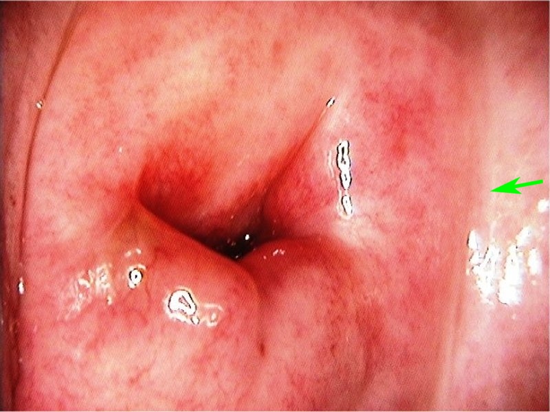
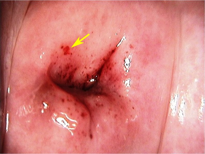
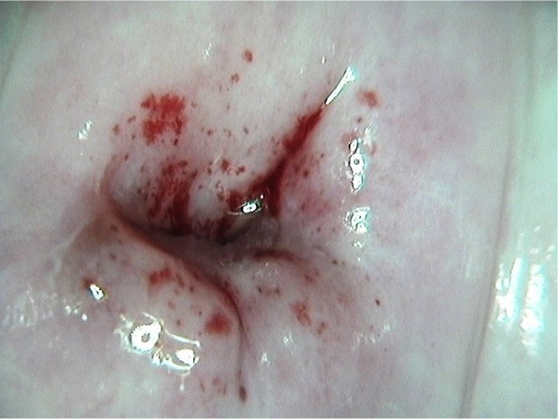
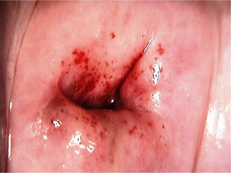
| Provisional diagnosis: | Type 2 transformation zone; normal cervix with atrophic change. |
| Management: | No further screening is required. |
| Histopathology: | Not done. |
| Comment: | The shallow fornix, pale colour of the epithelium, and squamocolumnar junction inside the canal indicate atrophic change. Petechial spots due to sub epithelial haemorrhage are more commonly seen in an atrophic cervix. Petechial spots should not be confused with coarse punctations. Petechial spots are flat, are of variable size, and can occur anywhere on the cervical and vaginal epithelium. Coarse punctations are always seen in the background of dense acetowhite epithelium and may be raised from the surface. |