|
Using Human Papillomavirus (HPV) detection tests for cervical cancer screening and managing HPV-positive women – a practical guide / Activity 2Anatomical considerations – Gross anatomy of female genital organs – Internal genital organs |   | .png)
Click on the pictures to magnify and display the legends
|
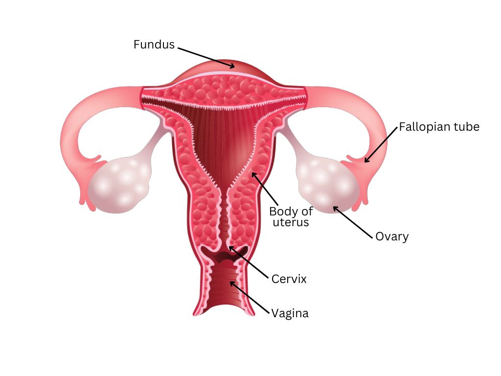
The components of the internal genital organs include the following:
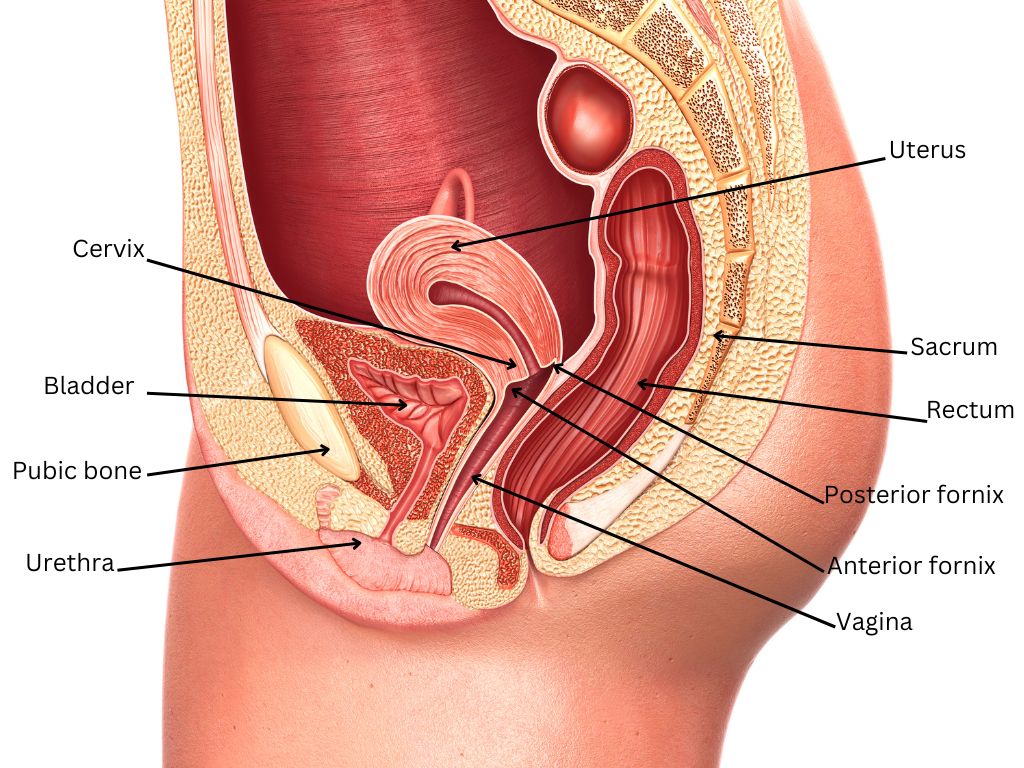
- Uterus: It is a hollow fibromuscular pear-shaped organ within which the fetus develops during pregnancy. It is located between the urinary bladder anteriorly and the rectum posteriorly. The parts of the uterus are:
- Fundus: The uppermost curved part that connects to the fallopian tubes at the corners.
- Body of the uterus: Starts from below the fundus and gradually narrows to form the lower part, known as the cervix.
- Cervix: The lower fibrous part that extends from the body of the uterus to open into the vagina. During a speculum examination, only part of the cervix is visible to the naked eye.
- Ovaries: These are small, egg-shaped glands located on either side of the uterus. They secrete hormones (estrogen and progesterone) that regulate the menstrual cycle and the fertility of a woman. The ovaries produce the ova, or egg cells, that are released during the menstrual cycles.
- Fallopian tubes: These are two narrow tubular structures attached to the corners of the fundus of the uterus. They allow the passage of ova released from the ovaries to the uterus. Fertilization of ova by sperm normally occurs within these tubes.
- Vagina: This is a fibromuscular tubular structure located between the urinary bladder and the urethra anteriorly and the rectum and the anal canal posteriorly. The cervix opens into the vagina at its upper end through the anterior wall. The vagina extends below to the introitus. When a woman is in the standing position, the vagina is directed upwards and posteriorly. When a woman is lying down on her back, the vagina is directed upwards and backwards.
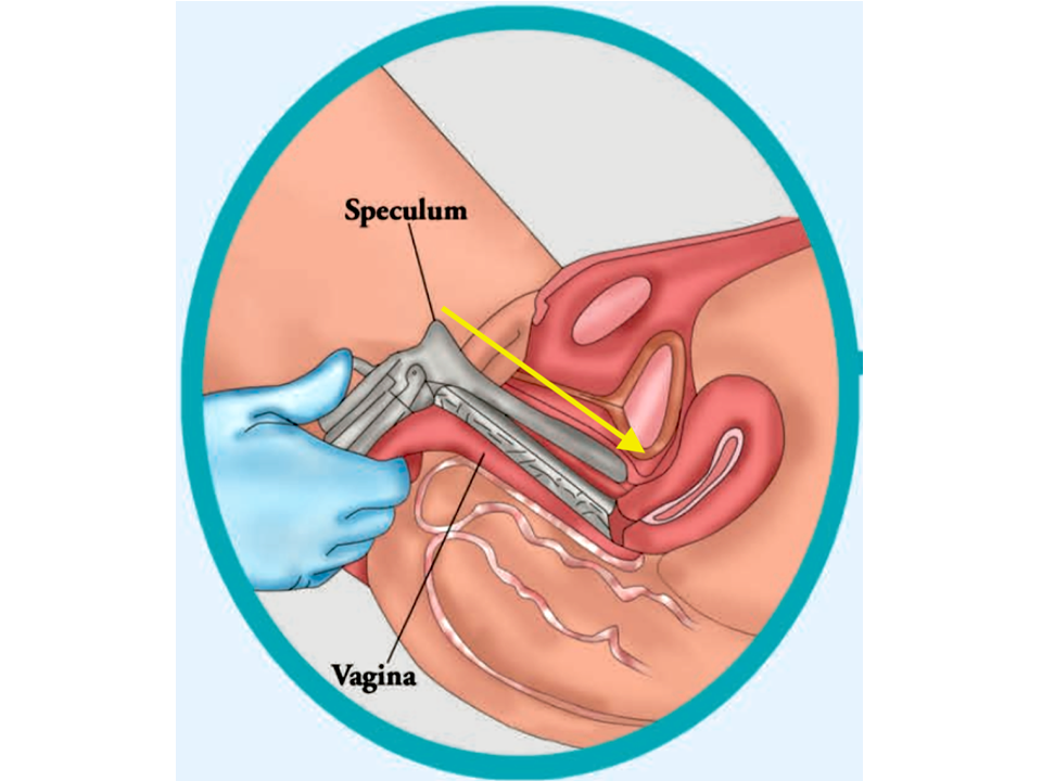
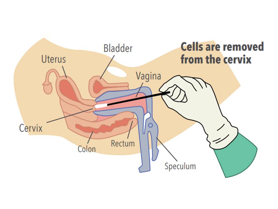
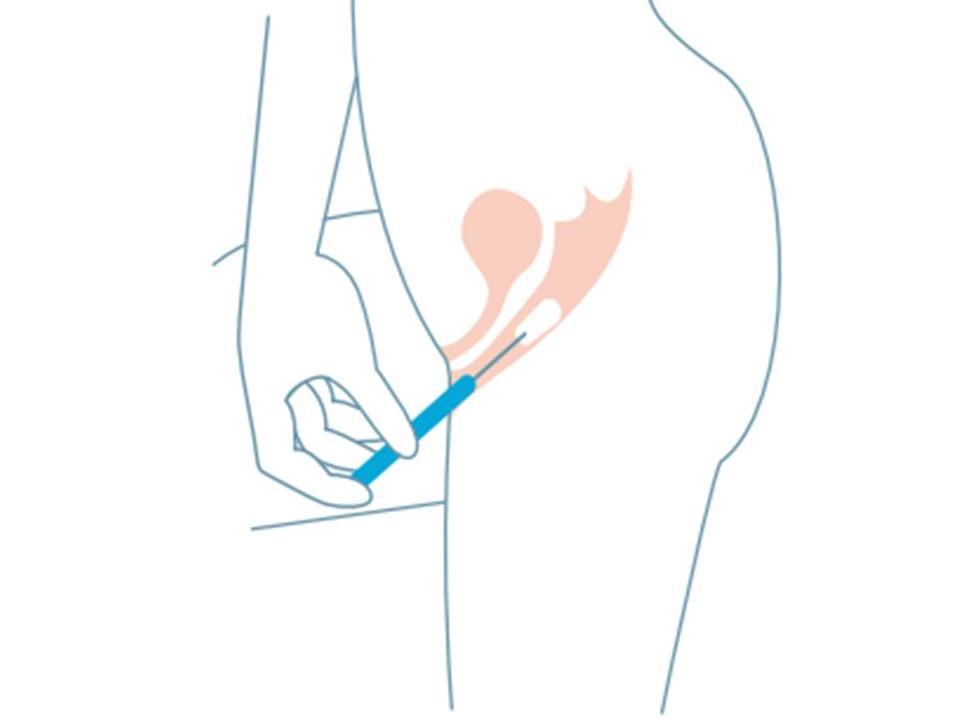
The direction of insertion of the speculum during the collection of a sample for HPV testing should be oriented towards the direction of the vagina, pointing towards the hollow of the sacrum, as shown in the diagram.
While self-collecting a vaginal specimen for HPV testing, a woman should direct the swab in the same direction (upwards and backwards while she is in a standing or squatting position).
|
The next section discusses the gross anatomy of the cervix.
|
| 