Using Human Papillomavirus (HPV) detection tests for cervical cancer screening and managing HPV-positive women – a practical guide / Activity 2
Anatomical considerations – Gross anatomy of the cervix | Click on the pictures to magnify and display the legends |
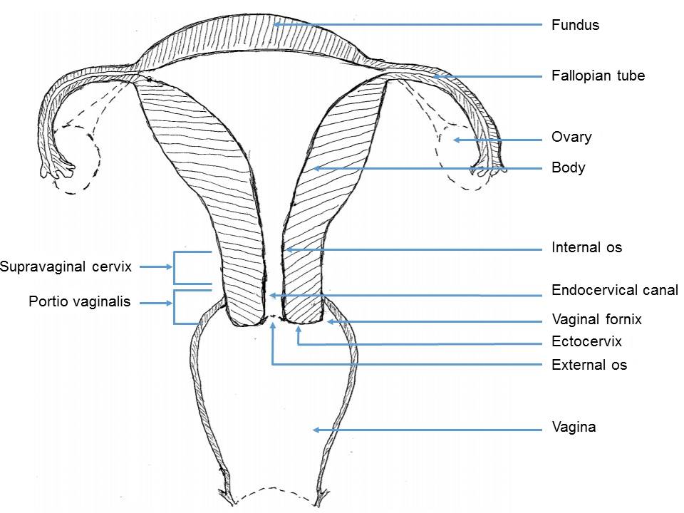
The cervix is the lower part of the uterus and is cylindrical. The length of the cervix is about 3–4 cm, and its diameter is about 2.5 cm. It has two parts:
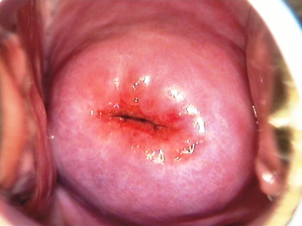
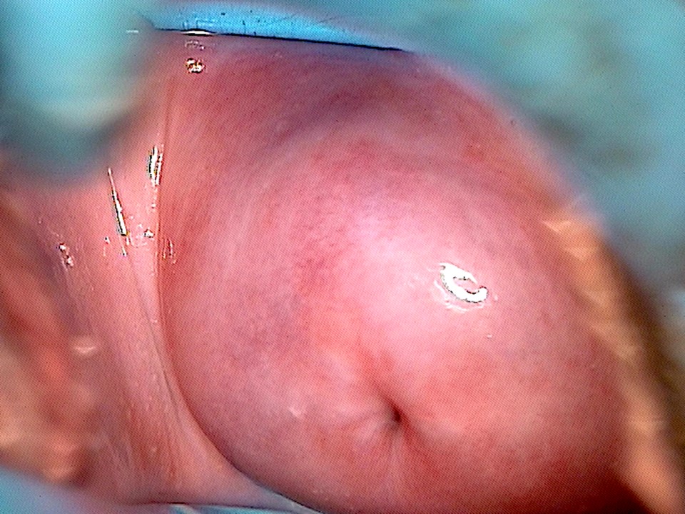
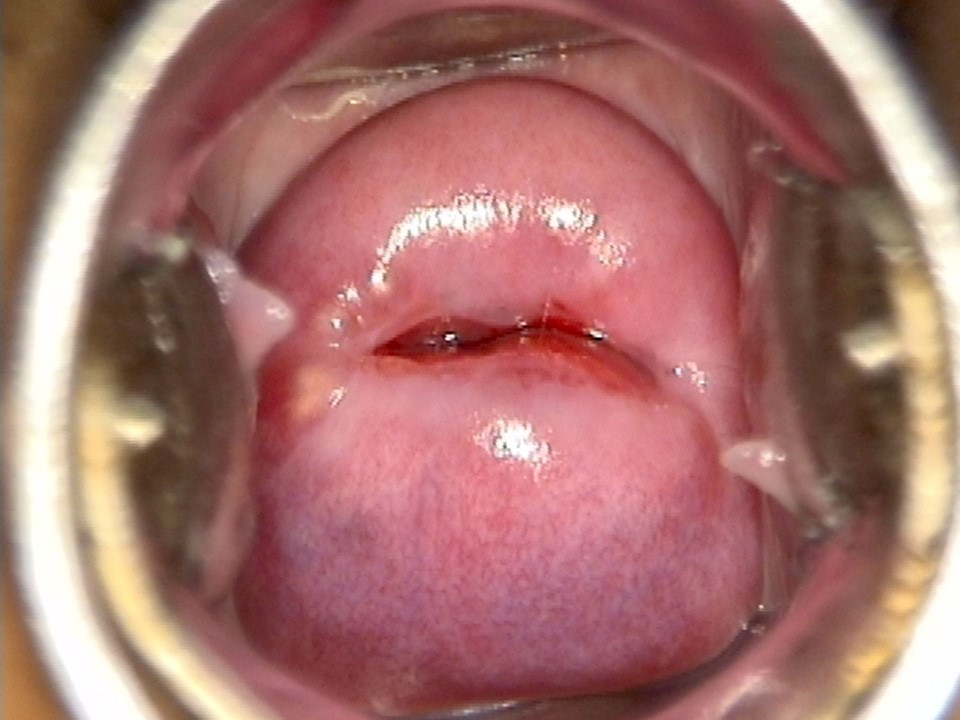
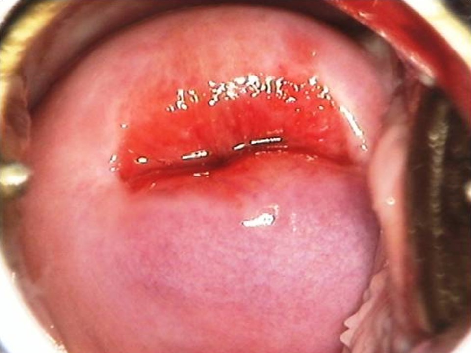
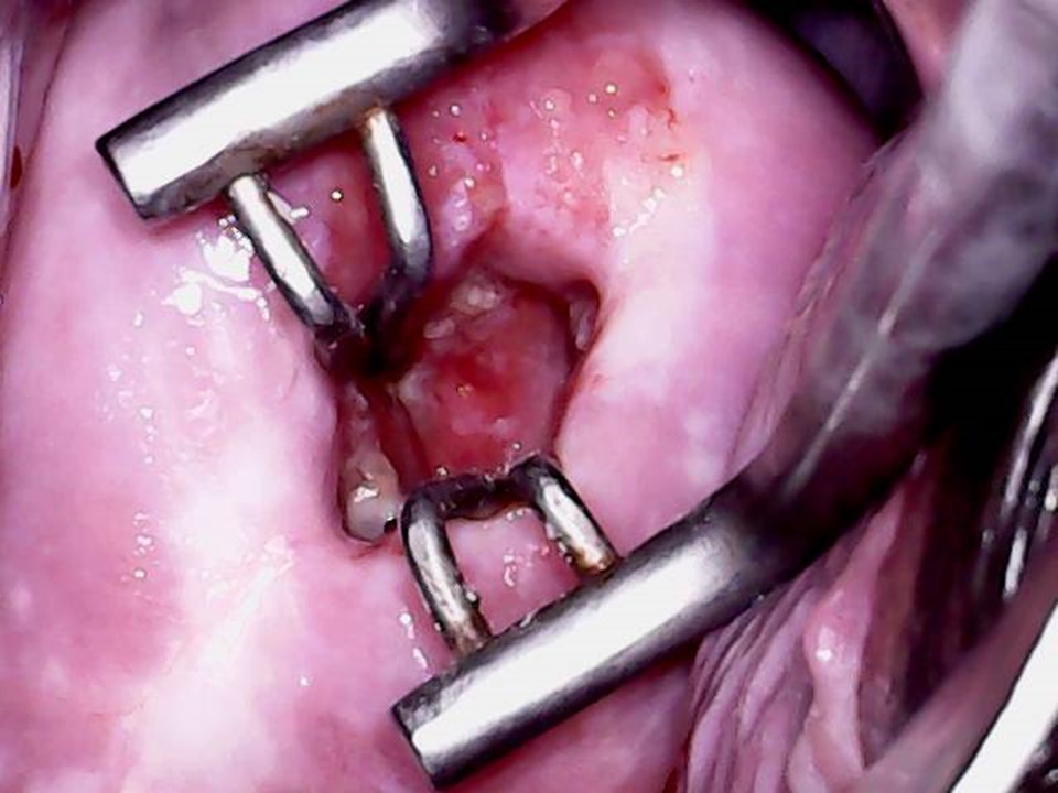
The part of the cervix that is visible during a speculum examination is the ectocervix. The cylindrical canal within the cervix is the endocervix. The endocervix and the ectocervix meet at the external os, which is an opening visible at the centre of the ectocervix. The endocervix joins the uterine cavity through the internal os. The ectocervix has an anterior lip and a posterior lip. In nulliparous women, the external os is small and round and resembles a pinhole. In parous women with history of vaginal childbirth the opening of the ectocervix is wide, it is transversely slit, and its shape resembles that of a fish’s mouth. The endocervical canal is a cylindrical space inside the cervix that opens into the vagina through the external os. The upper end of the endocervical canal opens into the body of the uterus through the internal os. The endocervix is lined with columnar epithelium and may not be visualized during a speculum examination of the cervix or during VIA. Endocervical visualization is possible after insertion of endocervical forceps (endocervical speculum). The next section discusses the microscopic anatomy of the cervix. |
