Atlas of visual inspection of the cervix with acetic acid for screening, triage, and assessment for treatment / Activity 1
Cervical epithelium – Squamous epithelium |
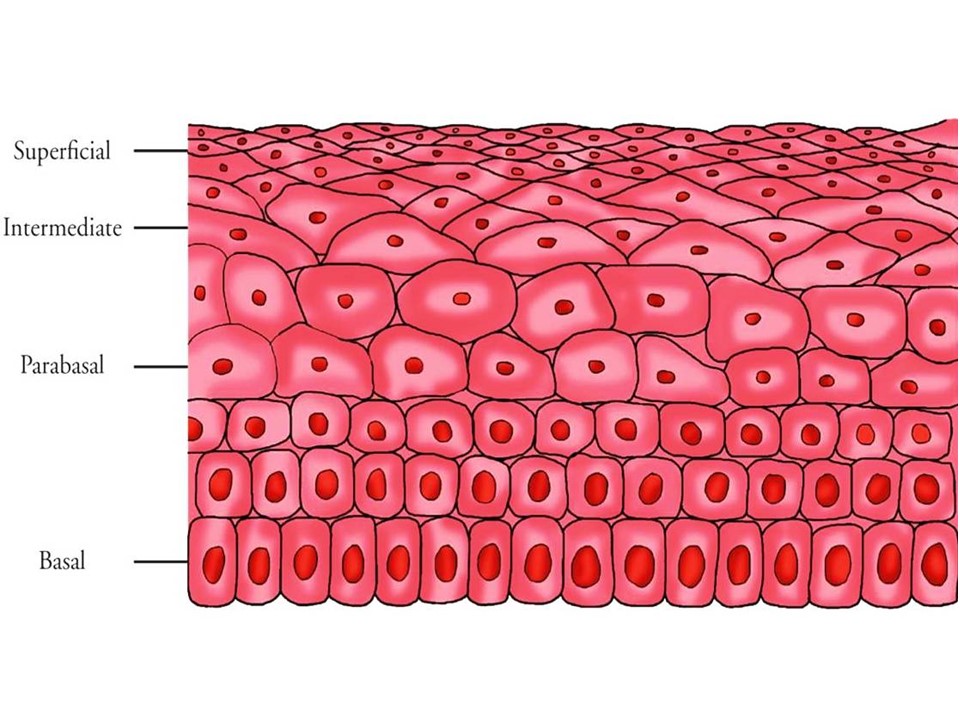
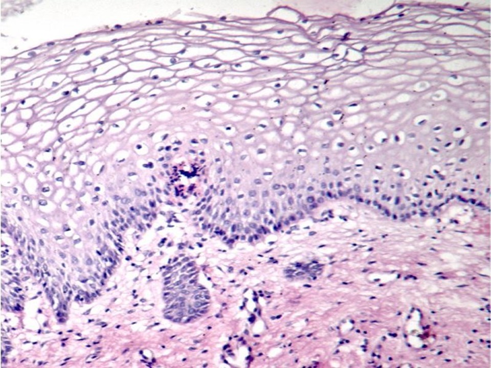
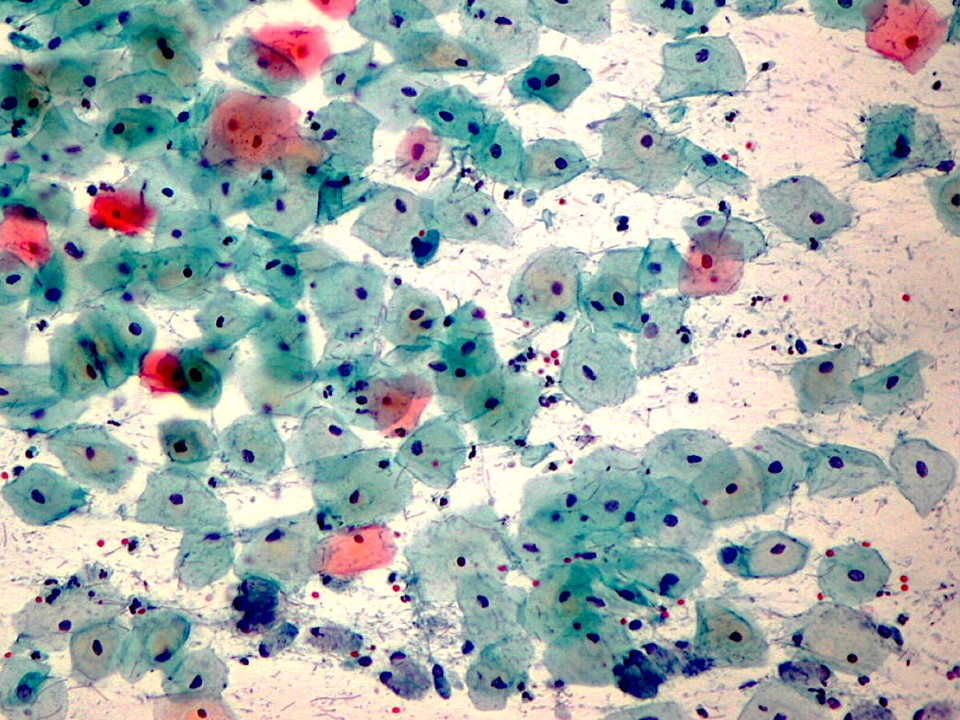
Naked-eye appearance of squamous epithelium Normal squamous epithelium appears smooth and covers the ectocervix. The epithelium is transparent. On naked-eye examination, the ectocervix appears pink because the underlying stroma is visible through the transparent epithelium. The cervical stroma appears red, because of the presence of abundant blood vessels. The transparent squamous epithelium acts as a colourless filter for the transmission of light. The squamous epithelium often meets the columnar epithelium around the external os. Microscopic features of squamous epithelium Squamous epithelium is composed of about 15–20 layers of squamous cells, neatly arranged in rows and separated from the underlying cervical stroma by a basement membrane. Such multilayered epithelium is known as stratified epithelium. Each cell has a nucleus at the centre and cytoplasm. The squamous epithelium is divided into the following four layers, starting from the lowest layer of cells:
The next section discusses the details of the columnar epithelium. |
Click on the pictures to magnify and display the legends


