Atlas of Colposcopy: Principles and Practice / Activity 6
Case |
High grade / CIN2 / CIN3
Go back to the list
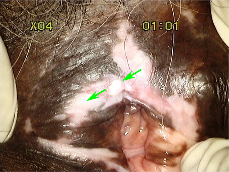 | |
| Vulva before acetic acid |
 | |
| Vulva after acetic acid |
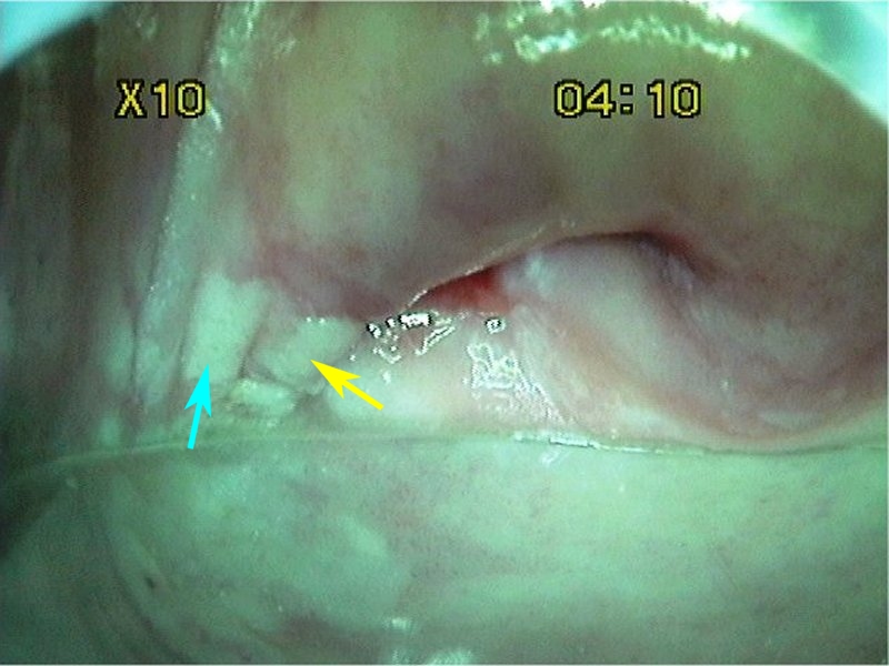 | |
| After acetic acid with green filter |
 | |
| After acetic acid with green filter |
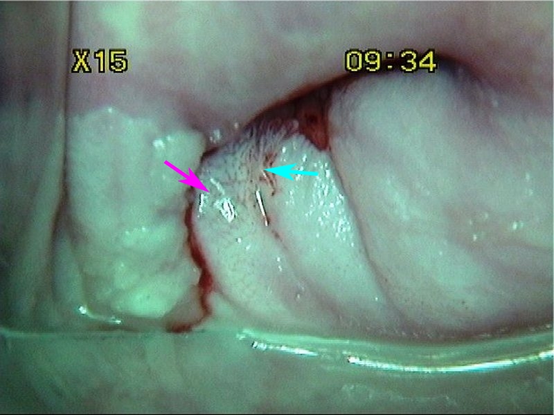 | |
| After acetic acid with green filter |
|
|
|
|
|
| |
Swede score:
| Nil or transparent | Thin, milky | Distinct, stearin | |
| Nil or diffuse | Sharp but irregular, jagged, satellites | Sharp and even, difference in level | |
| Fine, regular | Absent | Coarse or atypical vessels | |
| < 5 mm | 5-15 mm or 2 quadrants | >15 mm, 3-4 quadrants, or endocervically undefined | |
| Brown | Faintly or patchy yellow | Distinctly yellow |
Case Summary
| Provisional diagnosis: | Type 3 transformation zone; high-grade squamous intraepithelial lesion (HSIL) of the cervix, vagina, and vulva. |
| Management: | LLETZ (type 3 excision) and biopsies for vaginal and vulval lesions. |
| Histopathology: | Cervical histopathology was invasive squamous cell cancer, vaginal histopathology was HSIL-VAIN 3, and vulvar histopathology was HSIL-VIN 3. |
| Comment: | Multifocal neoplasias may be seen in the lower genital tract. The patient had a radical abdominal hysterectomy with bilateral pelvic lymphadenectomy and simple vulvectomy. |