Atlas of Colposcopy: Principles and Practice / Activity 6
Case |
Early and advanced cancers / Squamous cell cancer
Go back to the list
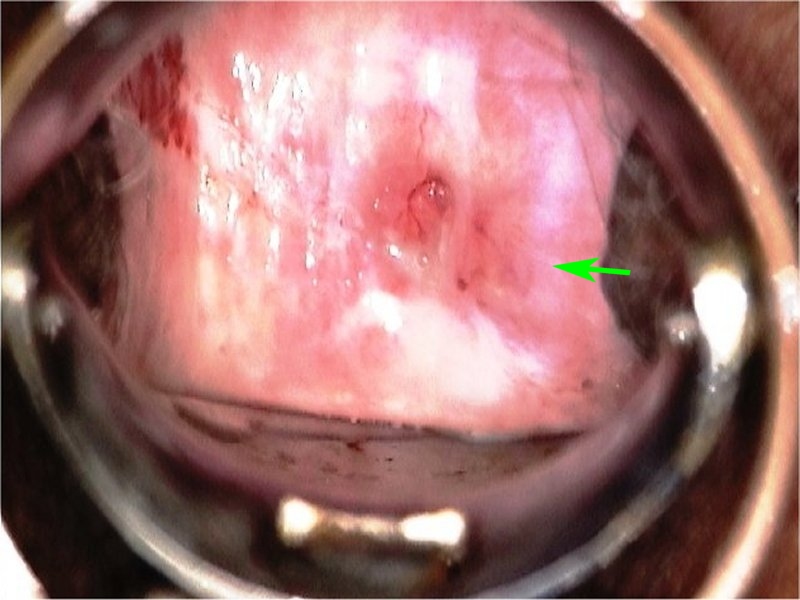 | |
| After normal saline |
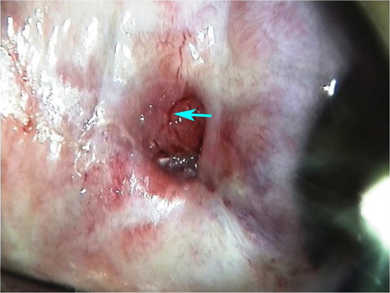 | |
| After normal saline with green filter |
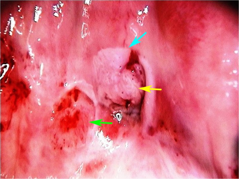 | |
| After acetic acid |
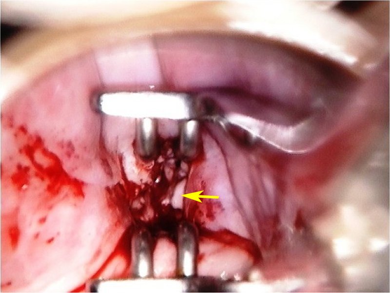 | |
| Visualization of the SCJ |
|
|
|
|
|
| |
Swede score:
| Nil or transparent | Thin, milky | Distinct, stearin | |
| Nil or diffuse | Sharp but irregular, jagged, satellites | Sharp and even, difference in level | |
| Fine, regular | Absent | Coarse or atypical vessels | |
| < 5 mm | 5-15 mm or 2 quadrants | >15 mm, 3-4 quadrants, or endocervically undefined | |
| Brown | Faintly or patchy yellow | Distinctly yellow |
Case Summary
| Provisional diagnosis: | Type 3 transformation zone; suspicion of invasive cancer likely to be squamous in nature. |
| Management: | Punch biopsy and endocervical curettage. |
| Histopathology: | Invasive squamous cell carcinoma. |
| Comment: | This case highlights the importance of endocervical assessment if the squamocolumnar junction is not fully visualized. The ectocervical lesion appeared as HSIL, and the cancer was confined to the canal. |