


Home / Training / Manuals / Atlas of breast cancer early detection / Cases
Atlas of breast cancer early detection
Go back to the list of case studies
.png) Click on the pictures to magnify and display the legends
Click on the pictures to magnify and display the legends
| Case number: | 136 |
| Age: | 38 |
| Clinical presentation: | Premenopausal woman with average risk of developing breast cancer presented with right breast lump noticed a few weeks ago. |
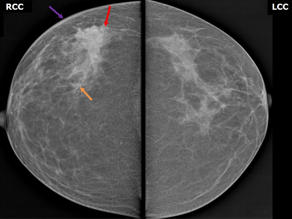
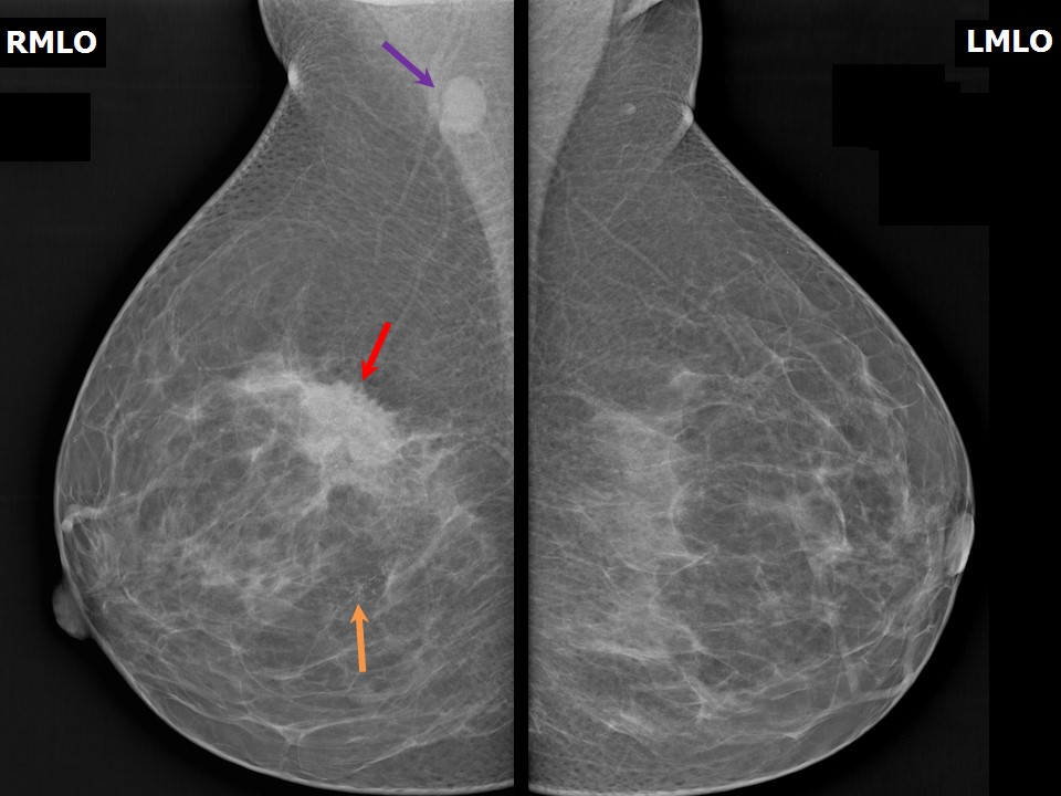
Mammography:
 |  |
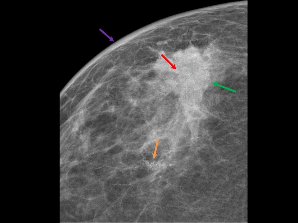 | 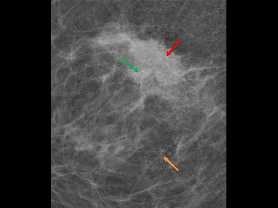 |
| Breast composition: | ACR category b (there are scattered areas of fibroglandular density) | Mammography features: |
| ‣ Location of the lesion: | Right breast, upper outer quadrant at 910 oclock, middle third, 6.0 cm from nipple and at 0.8 cm skin depth |
| ‣ Mass: | |
| • Number: | 1 |
| • Size: | 4.0 × 3.0 cm |
| • Shape: | Irregular |
| • Margins: | Spiculated |
| • Density: | High |
| ‣ Calcifications: | |
| • Typically benign: | None |
| • Suspicious: | Groups of fine pleomorphic microcalcifications within the mass and fine linear microcalcifications adjacent to the mass |
| • Distribution: | Grouped and linear distribution |
| ‣ Architectural distortion: | Present |
| ‣ Asymmetry: | None |
| ‣ Intramammary node: | None |
| ‣ Skin lesion: | None |
| ‣ Solitary dilated duct: | None |
| ‣ Associated features: | Axillary adenopathy and calcifications |
Ultrasound:
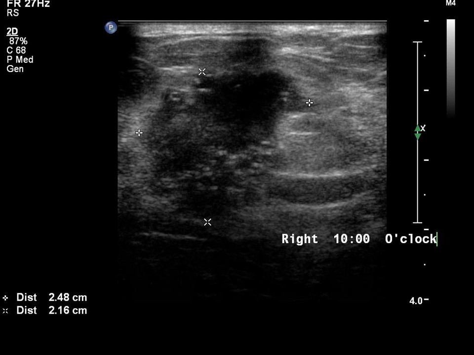 | 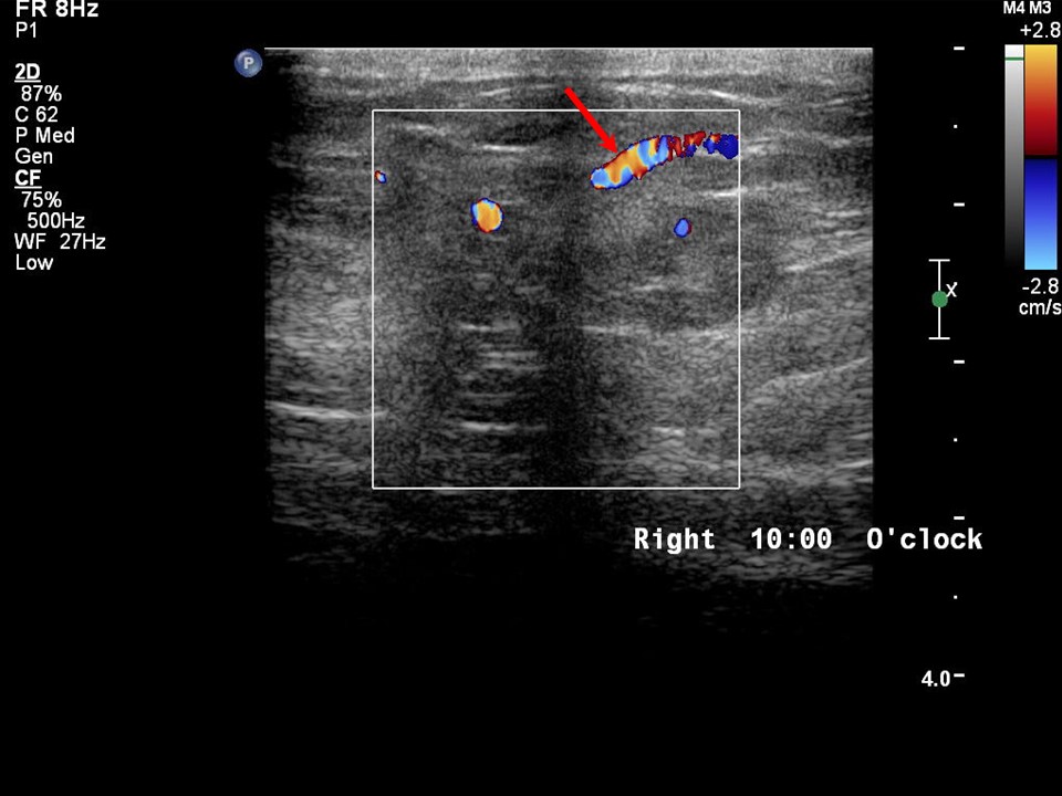 |
| Ultrasound features: Right breast, upper outer quadrant at 911 oclock | |
| ‣ Mass | |
| • Location: | Right breast, upper outer quadrant at 911 oclock |
| • Number: | 1 |
| • Size: | 2.5 × 2.2 cm |
| • Shape: | Irregular |
| • Orientation: | Not parallel |
| • Margins: | Indistinct and angular |
| • Echo pattern: | Heteroechoic |
| • Posterior features: | Combined pattern |
| ‣ Calcifications: | Grouped microcalcifications |
| ‣ Associated features: | Vascularity in mass and axillary lymphadenopathy |
| ‣ Special cases: | None |
BI-RADS:
BI-RADS Category: 5 (highly suggestive of malignancy)Further assessment:
Further assessment advised: Referral for cytologyCytology:
 |
| Cytology features: | |
| ‣ Type of sample: | FNAC |
| ‣ Site of biopsy: | |
| • Laterality: | Right |
| • Quadrant: | Upper outer |
| • Localization technique: | Palpation |
| • Nature of aspirate: | Thick whitish material |
| ‣ Cytological description: | Poorly cohesive clusters of malignant cells. Many single isolated malignant cells are seen |
| ‣ Reporting category: | Malignant |
| ‣ Diagnosis: | Carcinoma Breast |
| ‣ Comments: | None |
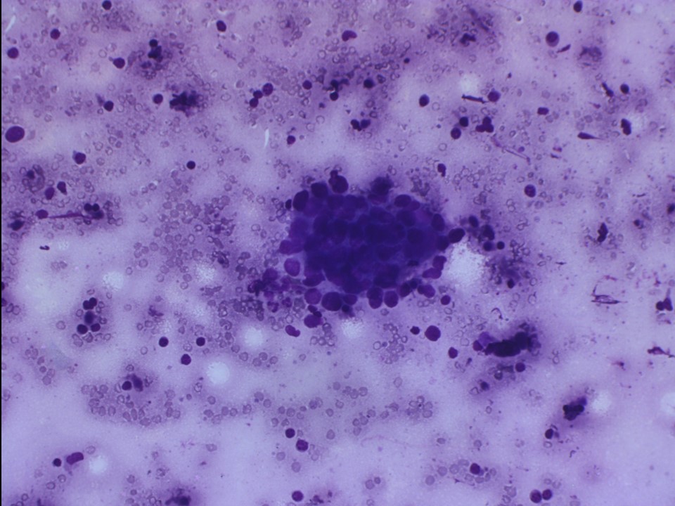 |
| Cytology features: | |
| ‣ Type of sample: | FNAC |
| ‣ Site of biopsy: | |
| • Laterality: | Right |
| • Quadrant: | Axillary node |
| • Localization technique: | Palpation |
| • Nature of aspirate: | Whitish |
| ‣ Cytological description: | Large malignant ductal epithelial cells seen in dyscohesive clusters on a background of lymphocytes |
| ‣ Reporting category: | Malignant |
| ‣ Diagnosis: | Metastases of breast carcinoma in axillary node |
| ‣ Comments: | None |
Histopathology:
Breast-conserving surgery
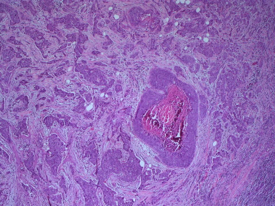 | 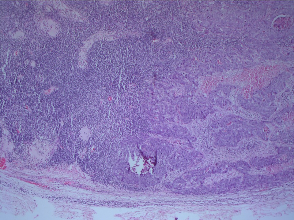 |
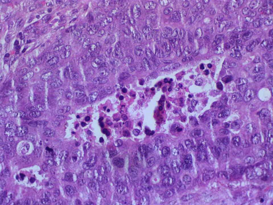 |
| Histopathology features: | |
| ‣ Specimen type: | Breast-conserving surgery |
| ‣ Laterality: | Right |
| ‣ Macroscopy: | Specimen (7.2 × 6.0 × 4.8 cm) and skin flap (4.0 × 1.0 cm). On serial sectioning, a solid greyish white tumour is identified (3.2 × 2.2 × 1.8 cm) |
| ‣ Histological type: | Invasive breast carcinoma |
| ‣ Histological grade: | Grade 3 (3 + 3 + 3 = 9) |
| ‣ Mitosis: | 22 |
| ‣ Maximum invasive tumour size: | 3.5 cm in greatest dimension |
| ‣ Lymph node status: | 1/17 |
| ‣ Peritumoural lymphovascular invasion: | Absent |
| ‣ DCIS/EIC: | DCIS of comedo and solid type high grade; EIC present |
| ‣ Margins: | Not involved |
| ‣ Pathological stage: | pT2N1 |
| ‣ Biomarkers: | |
| ‣ Comments: | Multiple calcific foci were seen in the necrotic areas in the malignancy |
Case summary:
| Premenopausal woman presented with right brPremenopausal woman presented with right breast lump. Diagnosed as right breast carcinoma with fine microcalcifications and overlying focal skin thickening, BI-RADS 5 on imaging, as breast carcinoma with axillary metastases on cytology, and as invasive breast carcinoma of no special type, pT2N1 on histopathology. east lump diagnosed as right breast carcinoma with fine microcalcifications and overlying focal skin thickening, BI-RADS category 5 on imaging, carcinoma breast with axillary metastases on cytology and Invasive Breast Carcinoma, NST , pT2N1 on histopathology. |
Learning points:
|