


Home / Training / Manuals / Atlas of breast cancer early detection / Cases
Atlas of breast cancer early detection
Go back to the list of case studies
.png) Click on the pictures to magnify and display the legends
Click on the pictures to magnify and display the legends
| Case number: | 086 |
| Age: | 43 |
| Clinical presentation: | Premenopausal woman under follow-up for fibrocystic changes in both breasts for more than 2 years. She had symptomatic breast nodularity in the left breast and presented with a left breast lump. On examination, a hard left breast lump was found. |
Mammography:
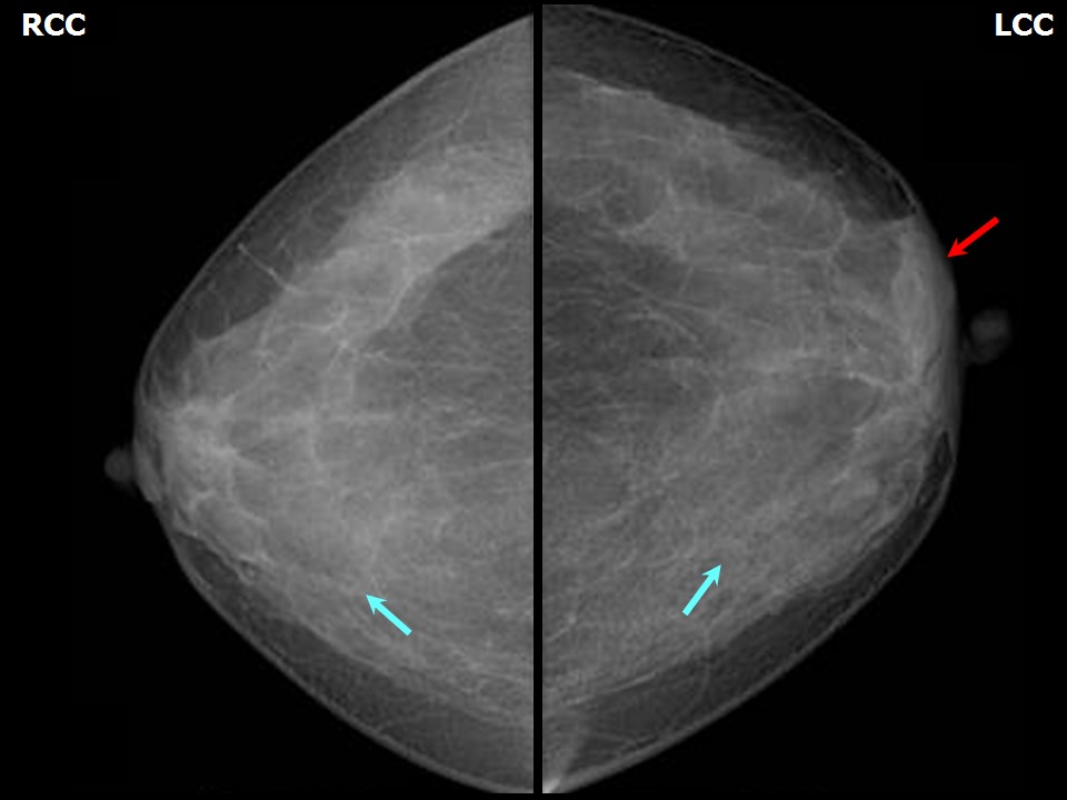 | 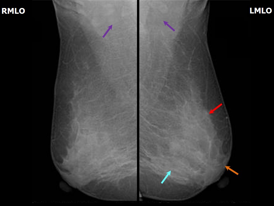 |
| Breast composition: | ACR category c (the breasts are heterogeneously dense, which may obscure small masses) | Mammography features: |
| ‣ Location of the lesion: | Left breast, upper inner quadrant at 11 oclock, middle third |
| ‣ Mass: | |
| • Number: | 1 |
| • Size: | Not measurable in dense breast parenchyma |
| • Shape: | Focal asymmetry |
| • Margins: | Indistinct |
| • Density: | Equal |
| ‣ Calcifications: | |
| • Typically benign: | None |
| • Suspicious: | None |
| • Distribution: | None |
| ‣ Architectural distortion: | None |
| ‣ Asymmetry: | Focal |
| ‣ Intramammary node: | None |
| ‣ Skin lesion: | None |
| ‣ Solitary dilated duct: | None |
| ‣ Associated features: | None |
Ultrasound:
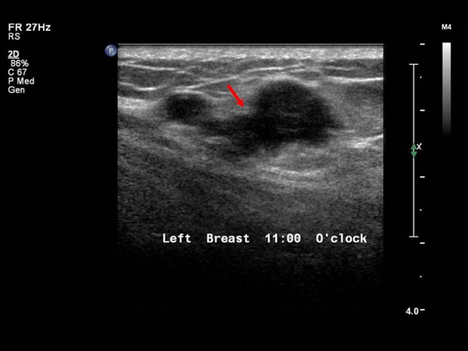 |  |
| Ultrasound features: Left breast, upper inner quadrant at 11 oclock | |
| ‣ Mass | |
| • Location: | Left breast, upper inner quadrant at 11 oclock |
| • Number: | 1 |
| • Size: | 2.6 × 1.0 cm |
| • Shape: | Irregular |
| • Orientation: | Not parallel |
| • Margins: | Angular |
| • Echo pattern: | Hypoechoic |
| • Posterior features: | No posterior features |
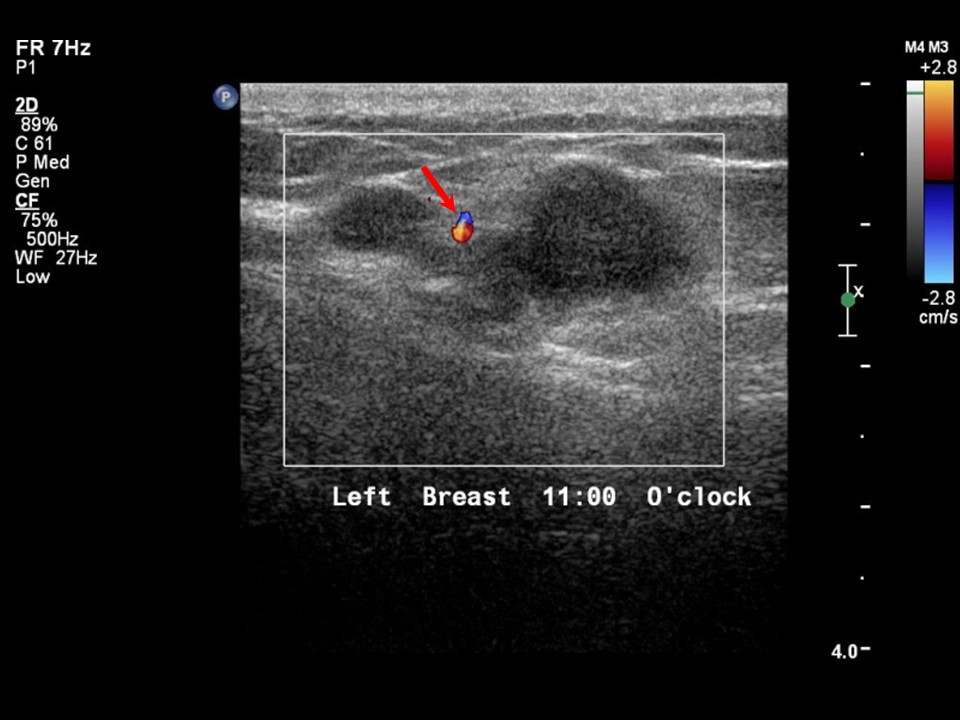
| ‣ Calcifications: | None |
| ‣ Associated features: | Internal vascularity |
| ‣ Special cases: | None |
BI-RADS:
BI-RADS Category: 4C (high suspicion for malignancy)Further assessment:
Further assessment advised: Referral for cytologyCytology:
 |
| Cytology features: | |
| ‣ Type of sample: | FNAC |
| ‣ Site of biopsy: | |
| • Laterality: | Left |
| • Quadrant: | Upper inner |
| • Localization technique: | Palpation |
| • Nature of aspirate: | Whitish |
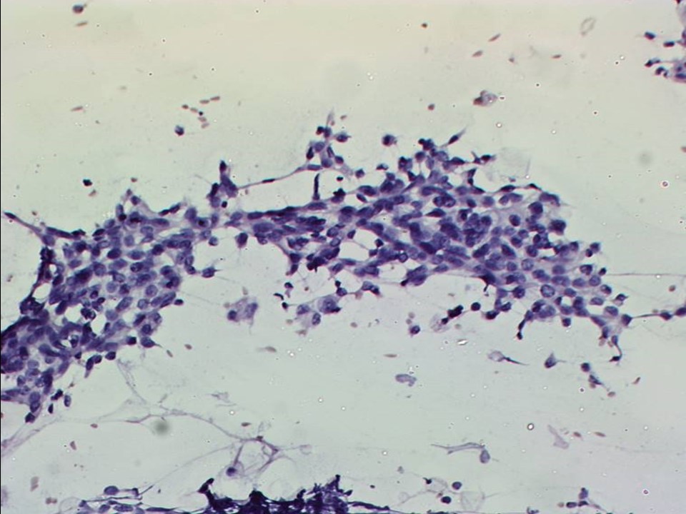
| ‣ Cytological description: | Smears are very cellular and reveal many dyscohesive clusters, sheets, and isolated atypical cells. The atypical cells have large, hyperchromatic and pleomorphic nuclei with prominent nucleoli. Many cells with bizarre nuclei are seen. Many lymphocytes are seen in the background |
| ‣ Reporting category: | Malignant |
| ‣ Diagnosis: | Carcinoma |
| ‣ Comments: | None |
Histopathology:
Breast-conserving surgery
 |
| Histopathology features: | |
| ‣ Specimen type: | Breast-conserving surgery |
| ‣ Laterality: | Left |
| ‣ Macroscopy: | Specimen (6.0 × 5.0 × 3.5 cm) and skin flap (5.0 × 3.0 cm). On serial sectioning, a tumour 2.3 × 1.5 × 1.8 cm is identified. Cut surface is greyish white. Tumour is located 2.0 cm from the skin, 0.8 cm from the posterior margin, 2.7 cm from the inferior margin, 2.0 cm from the medial margin, and 4.5 cm from the lateral margin. The superior margin is flush with the tumour. A small, very firm, whitish area is present flush with the inferior border 1.5 cm from the tumour. Adjacent breast shows a few areas of fibrosis. The superior margin was revised during surgery and was 2.5 cm from the tumour |
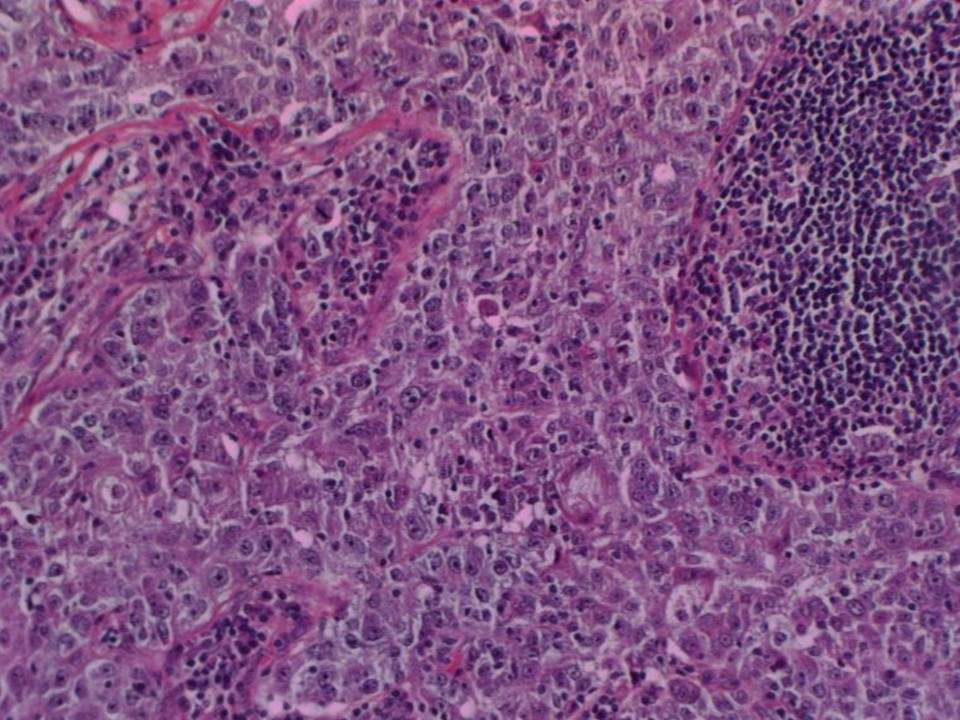
| ‣ Histological type: | Invasive carcinoma of no special type with medullary features |
| ‣ Histological grade: | Grade 3 (3 + 3 + 3 = 9) |
| ‣ Mitosis: | 20 |
| ‣ Maximum invasive tumour size: | 2.3 cm. The adjacent breast shows another small secondary tumour nodule 2 mm in diameter with poorly differentiated carcinoma in a syncytial pattern and in small cords surrounded by a dense infiltrate of lymphocytes |
| ‣ Lymph node status: | 0/14 |
| ‣ Peritumoural lymphovascular invasion: | |
| ‣ DCIS/EIC: | Absent |
| ‣ Margins: | Free of tumour; closest is posterior margin at 0.8 cm from the tumour |
| ‣ Pathological stage: | pT2(2)N0 |
| ‣ Biomarkers: | |
| ‣ Comments: | The section from the very firm area along the inferior border shows sclerosed stroma with normal lobules and ducts |
Case summary:
| Premenopausal woman presented with left breast lump. Diagnosed as focal asymmetry with areolar skin thickening in the left breast, BI-RADS 4C on imaging, as left breast carcinoma on cytology, and as invasive carcinoma of no special type with medullary features, pT2(2)N0 on histopathology. |
Learning points:
|