


Home / Training / Manuals / Atlas of breast cancer early detection / Cases
Atlas of breast cancer early detection
Go back to the list of case studies
.png) Click on the pictures to magnify and display the legends
Click on the pictures to magnify and display the legends
| Case number: | 063 |
| Age: | 39 |
| Clinical presentation: | Premenopausal woman with average risk of developing breast cancer presented with a rapidly growing left breast lump. On examination, she was found to have a large firm to hard lump in her left breast. |
Mammography:
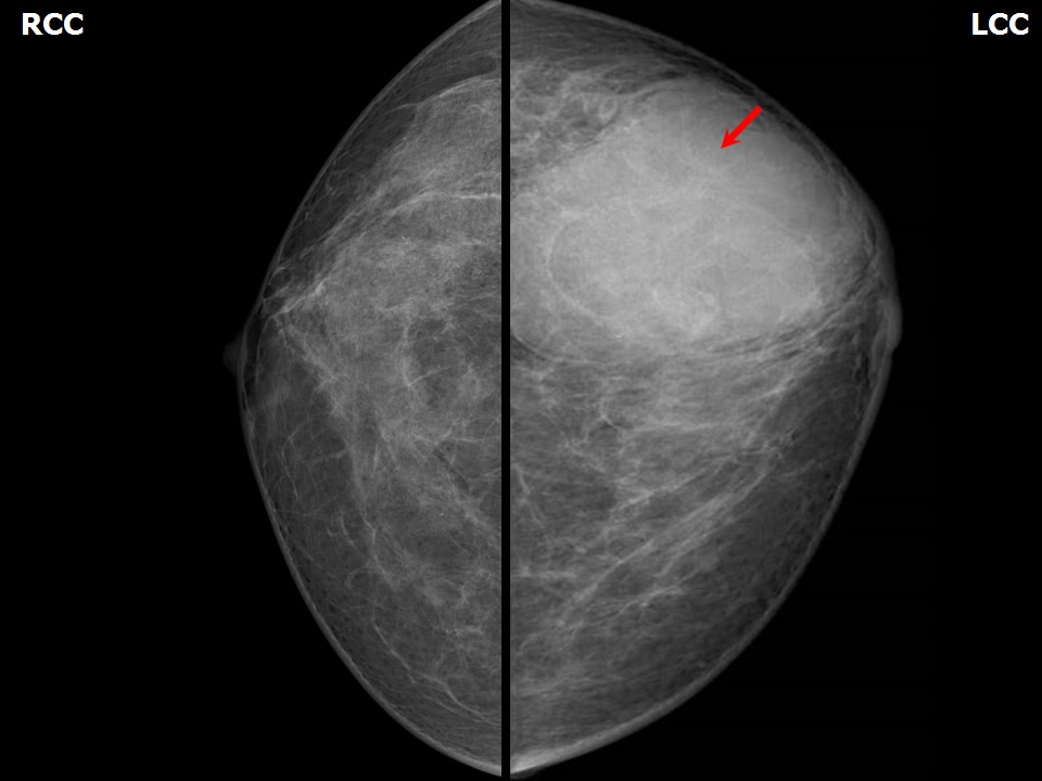 | 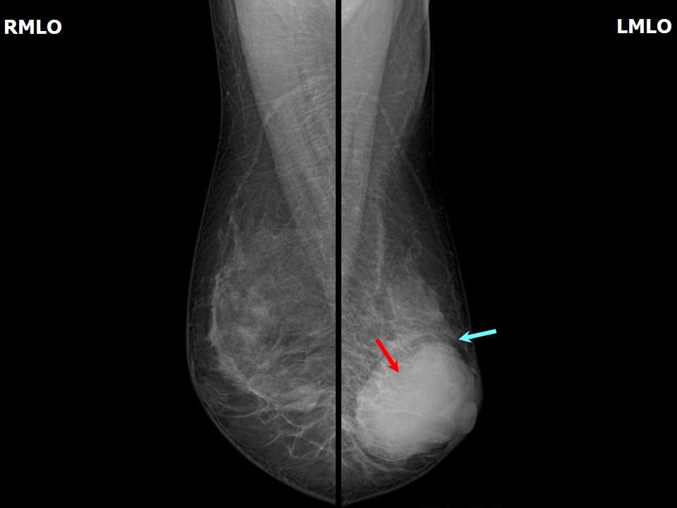 |
| Breast composition: | ACR category b (there are scattered areas of fibroglandular density) | Mammography features: |
| ‣ Location of the lesion: | Left breast, lower outer quadrant at 5 oclock, anterior, middle, and posterior thirds |
| ‣ Mass: | |
| • Number: | 1 |
| • Size: | 5.5 × 4.0 cm |
| • Shape: | Oval |
| • Margins: | Circumscribed |
| • Density: | High |
| ‣ Calcifications: | |
| • Typically benign: | None |
| • Suspicious: | None |
| • Distribution: | None |
| ‣ Architectural distortion: | None |
| ‣ Asymmetry: | None |
| ‣ Intramammary node: | None |
| ‣ Skin lesion: | None |
| ‣ Solitary dilated duct: | None |
| ‣ Associated features: | None |
Ultrasound:
 |  |
 |  |
 |
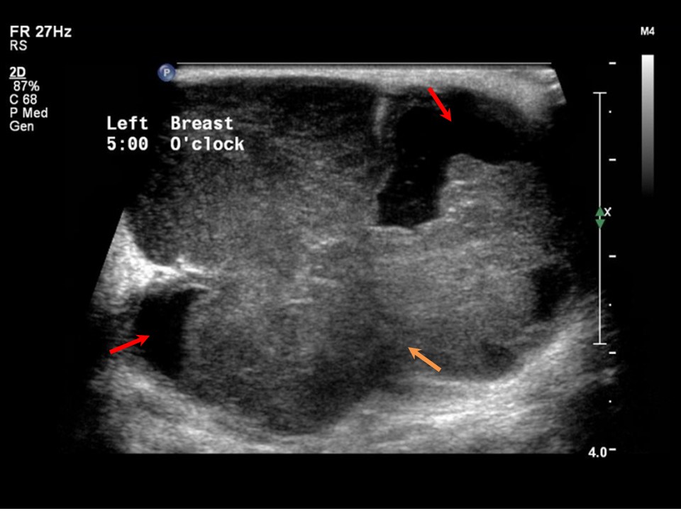
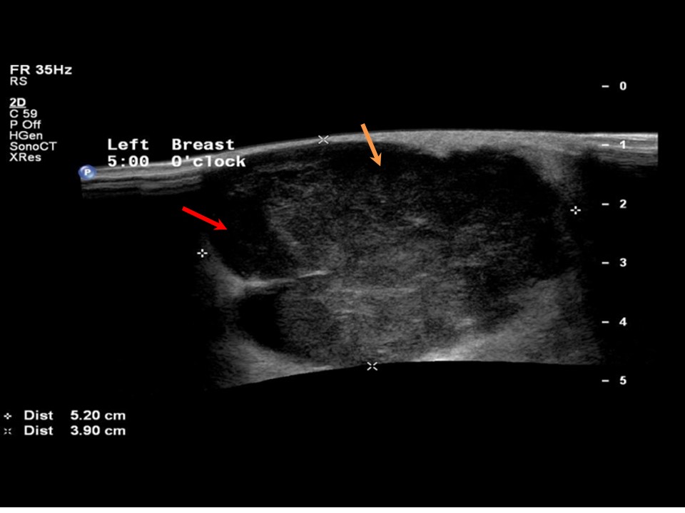
| Ultrasound features: Left breast, lower outer quadrant at 5 oclock | |
| ‣ Mass | |
| • Location: | Left breast, lower outer quadrant at 5 oclock |
| • Number: | 1 |
| • Size: | 5.2 × 4.0 cm |
| • Shape: | Irregular |
| • Orientation: | Parallel |
| • Margins: | Indistinct |
| • Echo pattern: | Heterogeneous with areas of breakdown |
| • Posterior features: | No posterior features |
| ‣ Calcifications: | None |
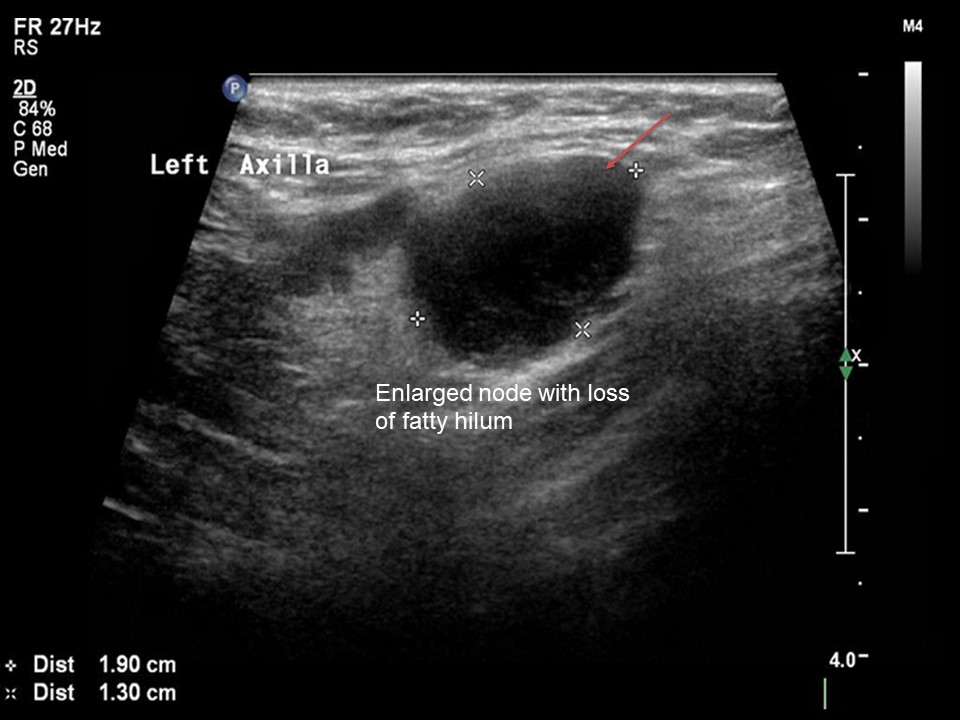
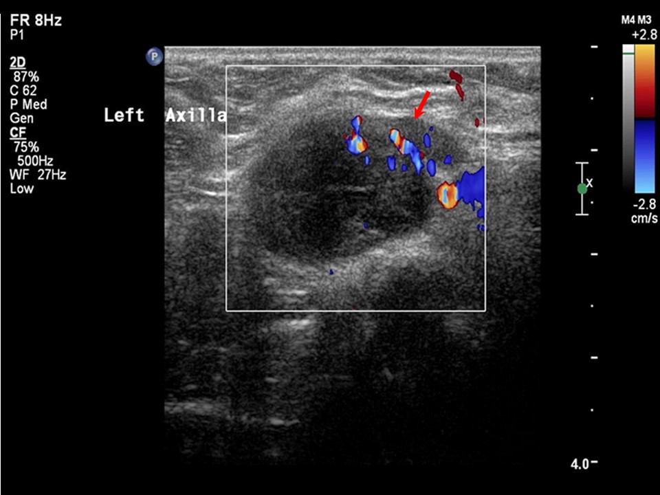
| ‣ Associated features: | Internal vascularity in mass. Enlarged lymph nodes (2.0 × 1.3 cm and 2.0 × 0.7 cm) with loss of central fatty hilum |
| ‣ Special cases: | None |
BI-RADS:
BI-RADS Category: 4C (high suspicion for malignancy)Further assessment:
Further assessment advised: Referral for cytologyCytology:
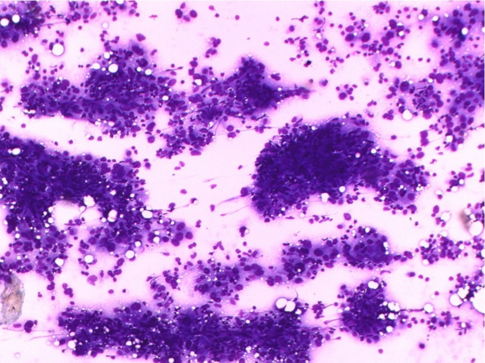 |
| Cytology features: | |
| ‣ Type of sample: | FNAC (solid lesion) |
| ‣ Site of biopsy: | |
| • Laterality: | Left |
| • Quadrant: | Lower outer |
| • Localization technique: | Palpation |
| • Nature of aspirate: | Whitish |
| ‣ Cytological description: | Highly pleomorphic malignant cells seen with lymphocytes in the background |
| ‣ Reporting category: | Malignant |
| ‣ Diagnosis: | Carcinoma high grade |
| ‣ Comments: | None |
Histopathology:
Breast-conserving surgery
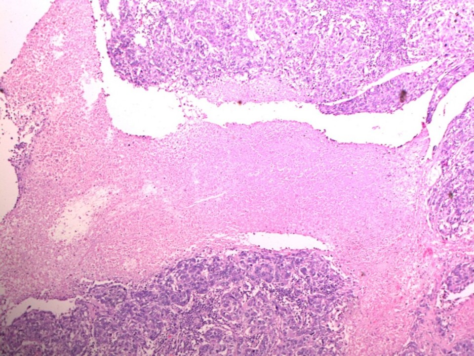 | 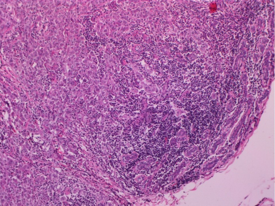 |
| Histopathology features: | |
| ‣ Specimen type: | Breast-conserving surgery |
| ‣ Laterality: | Left |
| ‣ Macroscopy: | Cut surface shows a large, greyish white, well-circumscribed tumour (5.5 × 5.0 × 4.0 cm) (T3) with a soft central necrotic area |
| ‣ Histological type: | Invasive breast carcinoma of no special type with medullary pattern |
| ‣ Histological grade: | Grade 3 (3 + 2 + 2 = 7) |
| ‣ Mitosis: | 15 |
| ‣ Maximum invasive tumour size: | 5.5 cm in greatest dimension |
| ‣ Lymph node status: | 1/24 |
| ‣ Peritumoural lymphovascular invasion: | Not identified |
| ‣ DCIS/EIC: | Not identified |
| ‣ Margins: | Free of tumour |
| ‣ Pathological stage: | pT3N1 |
| ‣ Biomarkers: | |
| ‣ Comments: | Extensive areas of tumour necrosis seen |
Case summary:
| Premenopausal woman presented with lump in the left breast. Diagnosed as circumscribed high-density mass with perilesional halo, with left axillary enlarged node of altered morphology, BI-RADS 4C on imaging, as breast carcinoma on cytology, and as invasive breast carcinoma of no specific type with medullary pattern, pT3N1 on histopathology. |
Learning points:
|