


Home / Training / Manuals / Atlas of breast cancer early detection / Cases
Atlas of breast cancer early detection
Go back to the list of case studies
.png) Click on the pictures to magnify and display the legends
Click on the pictures to magnify and display the legends
| Case number: | 045 |
| Age: | 67 |
| Clinical presentation: | Postmenopausal woman with average risk of developing breast cancer presented with a lump in the right breast. Examination revealed a 4 × 3 cm lump in the upper inner quadrant of the right breast. It was freely mobile and not fixed to the skin or chest wall. |
Mammography:
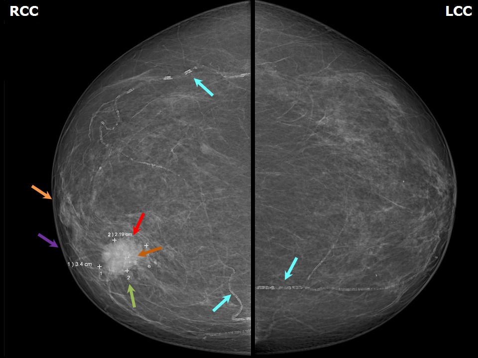 | 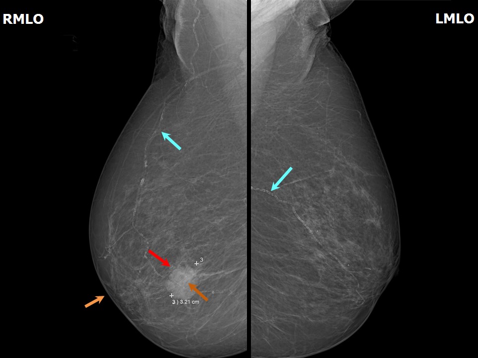 |
| Breast composition: | ACR category a (the breasts are almost entirely fatty) | Mammography features: |
| ‣ Location of the lesion: | Right breast, inner quadrant at 121 oclock, middle third |
| ‣ Mass: | |
| • Number: | 1 |
| • Size: | 3.4 × 3.2 × 2.2 cm |
| • Shape: | Irregular |
| • Margins: | Angular |
| • Density: | High |
| ‣ Calcifications: | |
| • Typically benign: | Vascular calcification |
| • Suspicious: | Fine pleomorphic |
| • Distribution: | In mass |
| ‣ Architectural distortion: | None |
| ‣ Asymmetry: | None |
| ‣ Intramammary node: | None |
| ‣ Skin lesion: | None |
| ‣ Solitary dilated duct: | None |
| ‣ Associated features: | Calcifications and axillary lymphadenopathy |
Ultrasound:
 |  |
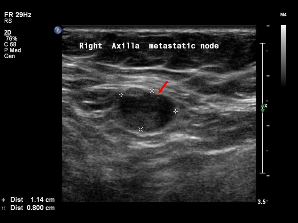 | 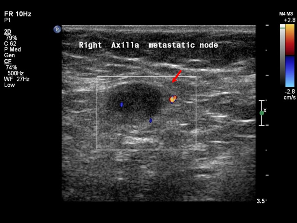 |
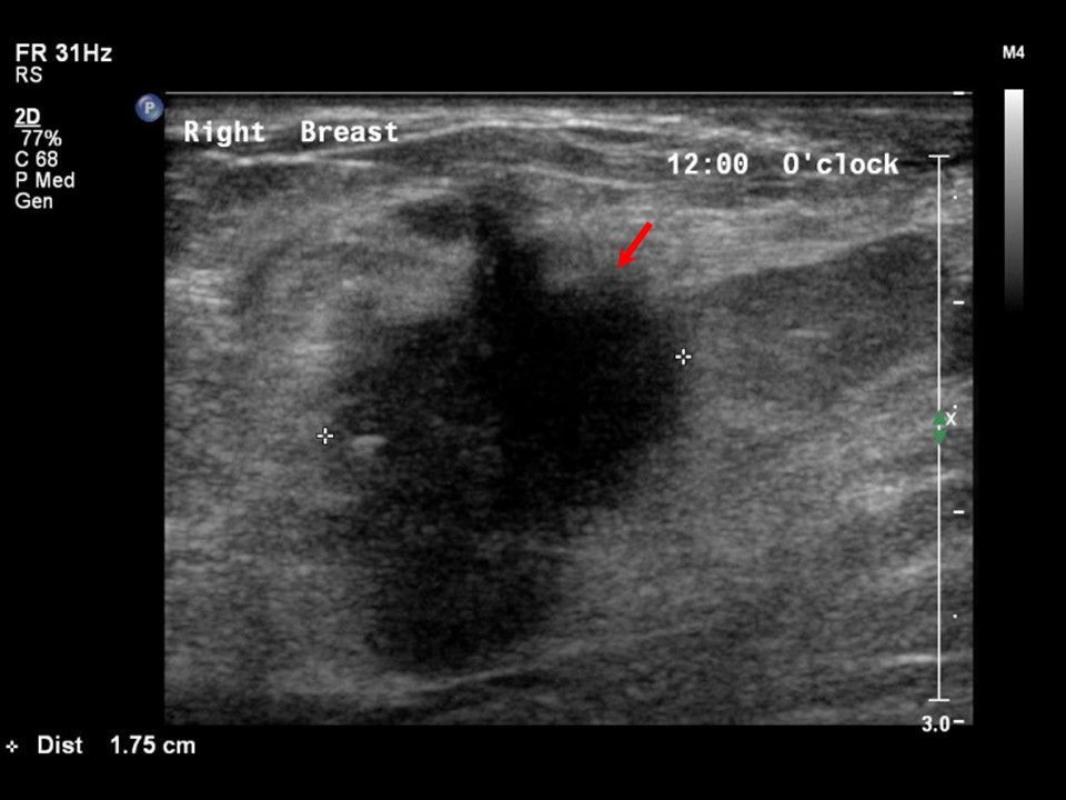
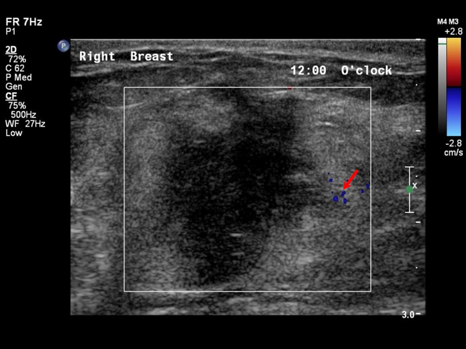
| Ultrasound features: Right breast, upper inner quadrant at 121 oclock | |
| ‣ Mass | |
| • Location: | Right breast, upper inner quadrant at 121 oclock |
| • Number: | 1 |
| • Size: | 3.2 × 2.0 cm |
| • Shape: | Irregular |
| • Orientation: | Not parallel |
| • Margins: | Angular |
| • Echo pattern: | Hypoechoic |
| • Posterior features: | Posterior shadowing |
| ‣ Calcifications: | Macrocalcifications in mass |
| ‣ Associated features: | Vessels in rim and axillary lymphadenopathy |
| ‣ Special cases: | None |
BI-RADS:
BI-RADS Category: 5 (highly suggestive of malignancy)Further assessment:
Further assessment advised: Referral for cytologyCytology:
 |
| Cytology features: | |
| ‣ Type of sample: | FNAC |
| ‣ Site of biopsy: | |
| • Laterality: | Right |
| • Quadrant: | |
| • Localization technique: | Palpation |
| • Nature of aspirate: | 0.5 mL of thick whitish material |
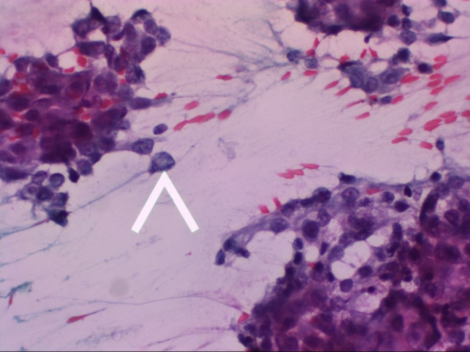
| ‣ Cytological description: | Smear shows tumour cells with overt malignant characteristics, such as dissociated cells (arrow), marked nuclear enlargement, nuclear hyperchromasia, and loosely cohesive cell clusters. (Pap; 400×) |
| ‣ Reporting category: | Malignant |
| ‣ Diagnosis: | Carcinoma |
| ‣ Comments: | None |
Histopathology:
BCS
 |
| Histopathology features: | |
| ‣ Specimen type: | BCS |
| ‣ Laterality: | Right |
| ‣ Macroscopy: | Right breast lumpectomy specimen (11.0 × 5.0 × 4.5 cm) with an elliptical flap of skin (5.0 × 1.5 cm). Cut surface shows a greyish white tumour (3.4 × 2.3 × 2.5 cm). The tumour is 2.5 cm below the skin, 3.0 cm from the base; 3.0 cm from the lateral margin, 2.3 cm from the medial margin, 2.0 cm from the superior margin, and 1.5 cm from the inferior margin. There is a smooth-walled cyst (2.5 cm in diameter) located inferior to the tumour |
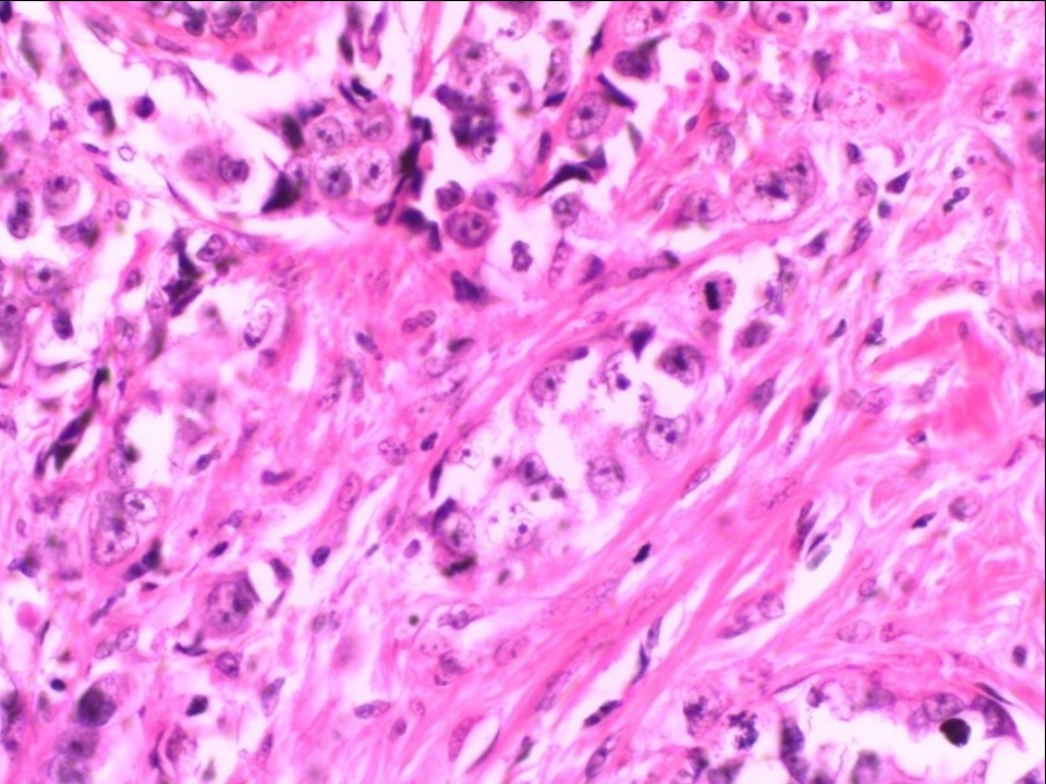
| ‣ Histological type: | Invasive breast carcinoma of no special type |
| ‣ Histological grade: | Grade 3 (3 + 3 + 2 = 8) |
| ‣ Mitosis: | 15 |
| ‣ Maximum invasive tumour size: | 3.4 cm in greatest dimension |
| ‣ Lymph node status: | 2/14 |
| ‣ Peritumoural lymphovascular invasion: | Absent |
| ‣ DCIS/EIC: | Comedo type DCIS of high grade; EIC absent |
| ‣ Margins: | Free of tumour |
| ‣ Pathological stage: | pT2N1 |
| ‣ Biomarkers: | |
| ‣ Comments: | Sections from the cyst inferior to the tumour show changes of fibrocystic change |
Case summary:
| Postmenopausal woman presented with right breast lump. Diagnosed as right breast carcinoma with coarse heterogeneous and fine microcalcifications within and nipple and skin retraction, BI-RADS 5 on imaging, as breast carcinoma on cytology, and as invasive breast carcinoma of no special type, pT2N1 on histopathology. |
Learning points:
|