


Home / Training / Manuals / Atlas of breast cancer early detection / Cases
Atlas of breast cancer early detection
Go back to the list of case studies
.png) Click on the pictures to magnify and display the legends
Click on the pictures to magnify and display the legends
| Case number: | 013 |
| Age: | 75 |
| Clinical presentation: | Postmenopausal woman with average risk of breast cancer presented with a painless right breast lump of long duration. The lump had gradually grown to the present size. Examination revealed a large firm lump (> 10 cm) in the right breast. |
Mammography:
 |  |
| Breast composition: | ACR category a (the breasts are almost entirely fatty) | Mammography features: |
| ‣ Location of the lesion: | Right breast, upper quadrants at 29 oclock, anterior, middle, and posterior thirds |
| ‣ Mass: | |
| • Number: | 1 |
| • Size: | 11.0 × 5.0 cm |
| • Shape: | Oval |
| • Margins: | Circumscribed |
| • Density: | Fat-containing |
| ‣ Calcifications: | |
| • Typically benign: | None |
| • Suspicious: | None |
| • Distribution: | None |
| ‣ Architectural distortion: | None |
| ‣ Asymmetry: | None |
| ‣ Intramammary node: | None |
| ‣ Skin lesion: | None |
| ‣ Solitary dilated duct: | None |
| ‣ Associated features: | None |
Ultrasound:
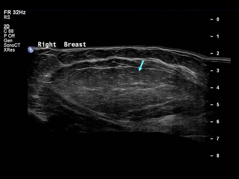 |
| Ultrasound features: Right breast, upper quadrants at 29 oclock | |
| ‣ Mass | |
| • Location: | Right breast, upper quadrants at 29 oclock |
| • Number: | 1 |
| • Size: | 10.5 × 4.5 cm |
| • Shape: | Oval |
| • Orientation: | Parallel |
| • Margins: | Circumscribed |
| • Echo pattern: | Isoechoic |
| • Posterior features: | No posterior features |
| ‣ Calcifications: | None |
| ‣ Associated features: | None |
| ‣ Special cases: | None |
BI-RADS:
BI-RADS Category: 2 (benign)Further assessment:
Further assessment advised: Referral for cytologyCytology:
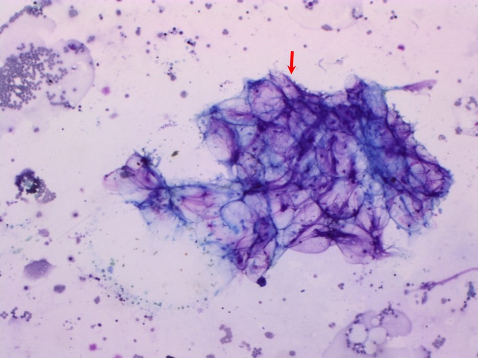 |
| Cytology features: | |
| ‣ Type of sample: | FNAC |
| ‣ Site of biopsy: | |
| • Laterality: | Right |
| • Quadrant: | Upper half |
| • Localization technique: | Palpation |
| • Nature of aspirate: | Greasy, yellowish material |
| ‣ Cytological description: | Smears show only adipose tissue fragments. Ductal epithelial cells not seen |
| ‣ Reporting category: | Benign |
| ‣ Diagnosis: | Only adipose tissue |
| ‣ Comments: | None |
Case summary:
| Postmenopausal woman presented with a large painless lump on her right breast of long duration. Diagnosed as lipoma, BI-RADS 2 on imaging and as benign adipose tissue on cytology. |
Learning points:
|