


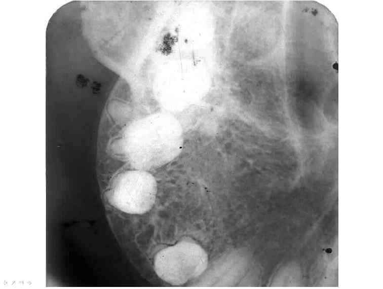
Figure 1: Central hemangioma of maxilla: Maxillary oblique occlusal view in which the normal architecture is replaced by coarse trabeculae. Expansion with tooth displacement is evident, but cortex is still present. The dark spaces between trabeculae are blood cavities.
25 avenue Tony Garnier CS 90627 69366, LYON CEDEX 07 France - Tel: +33 (0)4 72 73 84 85
© IARC 2024 - Terms of use - Privacy Policy.
© IARC 2024 - Terms of use - Privacy Policy.